 |
Manuscripts Collections are paper-generated collections that include correspondence, research notes, publications, scrapbooks, newspaper clippings, meeting minutes, departmental documents and transcripts. The Archives houses approximately 116 manuscript collections. Here are a few examples: |
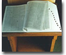
Photograph of Coy Cornelius Carpenter (From the Coy C. Carpenter Personal Collection)
Size: 5 cubic feet
Restrictions: Three cubic feet of the collection are available to the public upon completion of an interview with the Archivist. The remaining 2 cubic feet are restricted.
Preservation: Staples and paperclips have been removed. Materials have been placed in alkaline folders.
Number of boxes: 5
Provenance: Dr. Carpenter’s widow, Dorothy Carpenter, gave the collection to the Archives in 1979. In 1994 the material items were donated to the Archives by Dr. Harry Carpenter, Coy Carpenter’s son.
Biography: Coy Cornelius Carpenter, M.D. was born April 24, 1900 and died November 7, 1971. Dr. Carpenter graduated from Wake Forest College two-year medical school in 1922. He graduated from Syracuse Medical School in 1924 and received his certification in Atomic Pathology in 1936. After two postgraduate years at Syracuse as Instructor in Pathology and Clinical Medicine, he returned to North Carolina where he assumed the responsibilities for the administration of the medical school at Wake Forest College in Wake Forest, North Carolina in 1930. He became the founding dean of The Bowman Gray School of Medicine of Wake Forest University in Winston-Salem, North Carolina. He was dean from 1936 to 1963, when he became Vice-president for Medical Affairs, a post he continued until his retirement in 1967. When he was selected to fill this newly created post, he had been dean the longest of any medical school dean in the United States. During most of his term he also served as administrator, fundraiser, pathologist, and teacher. Dr. Carpenter formulated various programs, considered innovative at the time, in medical education. He established the Private Diagnostic Clinic and also served as Medical Director of North Carolina Baptist Hospital until 1953. As a Fulbright lecturer in Pathology, he spent 1953-1954 at Fouad University and Ibrahim University in Cairo, Egypt. Later he served as a consultant Medical Education for the International Cooperation Administration in South Vietnam. He was a Captain in the Medical Corp for the North Carolina National Guard from 1928 to 1938. His colleagues said of him, ". . . he was one of the true innovators in American Medical Education."
Scope of the Collection: The Coy C. Carpenter Collection consists of various materials ranging from personal correspondence to photographs to institutional brochures. The collection contains some materials generated from Wake Forest College in Wake Forest, however most of the materials are from the Bowman Gray School of Medicine at Wake Forest College in Winston-Salem.
Series Descriptions:
I. The series labeled correspondence has been arranged alphabetically and chronologically from 1933 to 1934. There are 133 files in this series.
II. The second series consists of the papers generated from a meeting held at Wake Forest College on Cancer. The renowned Joseph Bloodgood, cancer specialist, was a guest. This series contains correspondence, newspaper clippings, programs, and other documents related to the planning of the meeting.
III. The Newspaper clippings and publications are simply items Carpenter clipped that document various happenings at Wake Foes College in Wake Forest and Winston-Salem.
IV. The operational files for the Wake Forest College include such items as the first five-year report to the President, bank statements, deeds, statements for loans and grants, and solicitations for donations for a portrait of Dr. Thurman Kitchin.
V. This series consists of two speeches Carpenter gave on two different occasions.
VI. The material objects were given by Mrs. Carpenter and her son Harry Carpenter, M.D. They include an academic robe, the first doctor’s bag owned by C.C. Carpenter, passports, a journal, a pen, medical instruments, and a charm bracelet given to Dorothy by Coy.
VII. This entire series deals with the book written by Coy C. Carpenter, The Story of Medicine at Wake Forest University, and includes the original manuscript, correspondence relating to the book, and two letters by the editor.
VIII. The photographs are of a variety of subjects and size. Most all of them contain the likeness of Dr. Carpenter.
IX. Deans (restricted)
X. Memoirs (restricted)
Inventory: Series I: Correspondence|
Box 1: Correspondence A-Z, 1933-1934 Box 2: Correspondence A-Z, 1935-1939 |
Series II: North Carolina Medical Society Meeting on Cancer
|
Box 3: Letter Governor Hoey |
||
|
Committee on Cancer, NC Medical Society, WFU, October 18, 1933 Correspondence with J.W. Cox, Cancer Clinic, 1933 Cancer Clinic Joseph Colt Bloodgood American Society for the Prevention of Cancer, 1933 Letter from person with Cancer |
||
| Box 4: Early Days and move to Wake Forest | ||
|
At Wake Forest and Early Days in Winston-Salem New Campus Biblical Recorder and Program 1943 Medical Alumni News Article 1950s Trustee Actions on College Clippings – Growth and honors at the Medical School Dr. Thurman Kitchin Clippings Dr. Carpenter Clippings Clippings and Realia Clippings 1940s and 1950s Clippings 1940s Clippings 1948 Clippings 1950s Clippings 1950s Clippings 1960 Medical Alumni News 1967 Magazine Articles 1960s and 1970s Retirement and Portrait Presentation Clippings 1970 Clippings 1971 Wake Forest Magazine 1950 Wake Forest Magazine 1961 Wake Forest Magazine 1970 Wake Forest Magazine 1971 Reprints – Coy C. Carpenter North Carolina Medical Journal 1971 |
||
Series IV: Operational Files at Wake Forest College
|
Box 5: Stationery – Wake Forest College and Coy C. Carpenter |
||
|
Dr. Kitchin’s portrait – Solicitations for Donations Carpenter Medical Center Publications and copy of organization and policies handbook Deeds – contains photographs Wake Forest College
|
||
Series V: Speeches
|
Box 5: Speech Wingate College 1962 |
||
| Academic Transplant | ||
Series VI: Material Items
|
Box 6: Passports |
||
|
Journal Pen Medical Instruments Charm Bracelet Doctor’s Bag Scrapbook |
||
| Box 7: Academic Robe | ||
Series VII: The History of Medicine at Wake Forest
|
Box 8: Original Manuscript |
||
| Box 9: Book Reviews | ||
|
Book Reviews The History of Medicine at Wake Forest Letters from the editor Correspondence pertaining to book |
||
Series VIII: Photographs
Box 10 |
||
|
File 1: Architect’s Drawing 1950
File 2: Groundbreaking 1940
File 3: Carpenter with doctor 1940s
File 4: Hospital Hong Kong
|
||
Series IX: Deans (Restricted)
| Box 11 |
Series X: Memoirs (Restricted)
| Box 12 |
Series XI:
|
Back to the top.
Back to Archive Collections.
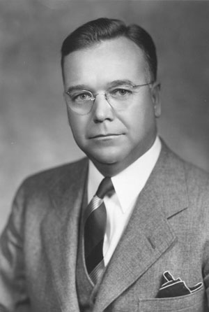
Engagement Photograph of Dorothy Carpenter, circa 1925 (From the Dorothy Carpenter Personal Collection)
Size: .6 cubic feet
Restrictions: The collection is available to the public upon completion of an interview with the Archives staff. Preservation: Staples and paperclips have been removed. Materials have been placed in alkaline folders. Number of boxes: 1 plus other materials Provenance: This collection was donated to the Archives by Dorothy Carpenter herself. Biography: (excerpted from Wake Forest Magazine, April 1989, article by Mary Dalton)For decades, Dorothy Carpenter’s life has been entwined with the medical school. Her late husband, Dr. Coy C. Carpenter, was professor of pathology from 1926-1970, dean of the medical school from 1936-1963, and vice president for medical affairs from 1963-1967. He was instrumental in the medical school’s move to Winston-Salem and the development of the medical center. Throughout it all, Dorothy Carpenter was more than a helpmate – she was half of a team.
Dorothy Mitten Carpenter was born and reared in a small town in Delaware; she met Coy Carpenter when she was a student at Syracuse University and he was in medical school there. Wake Forest, North Carolina felt something like home when the couple moved there. "I entered into the life and activities of the college town to the extent that I directed three plays for the Drama Club of Wake Forest," said Mrs. {Carpenter}. "It was such a small town that if you were having a party and wanted lettuce, you had to order it a week in advance from 17 miles away."
"Life became a merry-go-round with a husband who had one project after another, but it was fun and exciting," she remarked. She says few people remember that it was her late husband who conceived the idea of moving the medical school to Winston-Salem. Dr. Carpenter was found of saying about the school that, "We took a shoestring and built a shoe around it." Mrs. {Carpenter} recalls many nights when her late husband lay awake worrying about finding money to pay faculty salaries. Somehow the money always came through, and the Bowman Gray School of Medicine began to grow. Mrs. {Carpenter} is as responsible in her own way for the development and success of the school as was Dr. Carpenter. She contributed the intangibles. The medical school moved to Winston-Salem with a faculty of seven. Mrs. {Carpenter} became a one-woman relocation and public relations firm. She was a real estate agent, babysitter, tour guide."My husband was building a school. In the selection of faculty members, my job was to sell Winston-Salem. I showed them the city, the homes, the schools; I got them a cook or anything else they wanted to make them happy. Some stayed with us—some just dined with us. At that time, the Robert E. Lee was the only hotel in town," she said.
In the meantime, Mrs. {Carpenter} had become the walking historian of the Bowman Gray School of Medicine. She kept a scrapbook and managed to stay on top of everything that was happening. In her "free time," Mrs. {Carpenter} served on the Red Cross Board, the YWCA Board, and the Board of Visitors of Peace College (where she went to boarding school before college.) As the unofficial Bowman Gray cheerleader and moral booster, Mrs. {Carpenter} organized the faculty wives into what is now called the Medical Center Auxiliary. She wanted to provide services to the medical center and a forum for the faculty to get to know one another. As usual, Mrs. {Carpenter} was working to be sure those around her felt comfortable and included. Mrs. {Carpenter} now confines most of her activities at Bowman Gray to the Coy C. Carpenter Library and the Dorothy Carpenter Medical Archives. The dedication of the archives last year was an appropriate recognition of the woman who for so many years was the archives and continues to be inexorably linked to the medial school’s success.She is especially proud of the library – with just cause. The Carpenter Library is one of the strengths of the Bowman Gray School of Medicine. There are over 135,000 volumes, over 3,000 journal titles, completely computerized services, and 10 professional librarians who oversee it.
When she talks about how she feels about Bowman Gray, Mrs. {Carpenter} smiles and admits that the school has been such an integral part of her life that she really does not know anything else. She counts among her finest hours her acceptance as an honorary member of the Bowman Gray Alumni Association in 1986. The alumni had indeed recognized one of their own
Scope of the Collection: This collection reflects, in part, the history of the move of Wake Forest College from Wake Forest and the lives of the Carpenters as they are involved with the medical school. The collection was donated by Dorothy Carpenter.
Inventory:|
3 Scrapbooks:
Folders: File 1: Correspondence, of Dorothy Carpenter File 2: Honolulu Newspaper D. Carpenter File 3: Coy C. Carpenter’s death File 4: Award to Dorothy, June 10, 1948 File 5: Articles, Clippings, and Publications File 6: Miscellaneous |
Back to the top.
Back to Archive Collections.
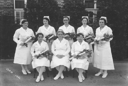
The North Carolina Baptist Hospital School of Nursing Class of 1932. Pictured in the back row from left to right: Ruby Jenkins Stainback, Pauline Binkley Jacobson, Thelma Lloyd Stroud, Elsie Lee Dall, Mary Taylor Maddrey. Front row from left to right: Lucille Cain Hartman, Edna L. Heinzerling, Mabel Meece (From the North Carolina Baptist Hospital School of Nursing Collection)
Go to NCBH School of Nursing Online Exhibit
Size: 5 cubic feet
Restrictions: The collection is available to the public upon completion of an interview with the Archives staff.
Preservation: Staples and paperclips have been removed. Materials have been placed in alkaline folders. Textiles have been wrapped in alkaline tissue and placed in alkaline boxes.
Number of Boxes: 9
Provenance: Varies with each series
History:
*Click for a PDF file of Ms. Heinzerling's 1960 North Carolina Baptist Hospital and School of Nursing history*
"In conjunction with the opening of the 88-bed, 22 bassinet North Carolina Baptist Hospital, the Baptist Hospital School of Nursing opened in 1923. The first class of 15 students was under the leadership of Miss Edna Heinzerling, RN, Director of the School of Nursing, as well as Director of Nurses for the Hospital."
"The education of the first student nurses included instruction by physicians and nurses, as well as working long hours in the hospital. The standard nursing attire included a long-sleeved blue uniform with a white apron, bib, collar, and cuffs. Black shoes and stockings were later changed to white. The white cap with black band became a standard part of the student uniform. The banding ceremony, at which the graduate nurse received a wide black velvet band, was a hallmark to which student nurses aspired. The first commencement exercise, held on May 25, 1926 at the First Baptist Church, graduated 10 students. Each graduate received a diploma and a North Carolina Baptist Hospital pin. This black and gold pin was embossed with the Florence Nightingale lamp. Ms. Heinzerling resigned in 1931 and was succeeded by Miss Lillian Anderson, RN. Ms. Heinzerling returned to her position in 1932, but due to illness was forced to resign again in 1936. Ms. Ruth Council, RN succeeded Ms. Heinzerling. During her tenure, the nursing school continued to increase enrollments, and in 1936, 18 living quarters were added to Blanche Barrus Nurses’ Home."
"In 1939 Ms. Council resigned, and Mrs. Leatha Smithdeal, RN, a Baptist graduate was appointed Acting Director of Nursing. Under her leadership, both graduate nurses and nursing students celebrated a dramatic change in their schedules. Their workweek was reduced from ten hours per day, seven days per week to eight hours per day. Mrs. Smithdeal was successful in constructing a six day work week for graduate nurses. She pioneered Public Health Nursing as a curriculum elective and established pediatric affiliations with Children’s Hospital of the University of Pittsburgh." "Just cause for celebration came in 1941 with the return of Ms. Heinzerling. During her absence, she had remained professionally involved by editing the historical text, The History of Nursing in North Carolina by Mary Lewis Wyche.""The Year 1941 also marked expansion at Baptist Hospital. The hospital increased its capacity to over 270 beds and 50 bassinets, and opened the first hospital pharmacy. This new service, under the direction of Mr. E.W. Rollins, Chief Pharmacist, replaced a small Drug Room where the Director of Nurses and her assistants acquired medications which they dispensed to patients throughout the hospital."
"Bowman Gray School of Medicine published its first yearbook, The Gray Matter, in 1942, and one year later The School of Nursing published its first yearbook, The Lamp. These yearbooks were combined in 1944 when seniors of both medical and nursing schools voted to publish one joint yearbook, The Gray and White Matter."
"The formation of the North Carolina Student Nurses’ Association and the election of Dorothy Inscore O’Briant, SN as the first president of the association marked a historic occasion in 1951. Ms. O’Briant was characterized by nursing instructors as an individual with tremendous leadership ability who served as a role model to which other students aspired."
"Mrs. Joyce Warren, RN, AB, BS succeeded Ms. Heinzerling as Director of Nursing and Director of the School of Nursing in 1952. Under her guidance in 1956, the School of Nursing became the largest diploma school in North Carolina. National accreditation was achieved in 1959, recognizing the excellence of the education program and placing the school among the elite few."
"When Ms. Warren retired in 1973, Mrs. Gwen Andrews, RN, MSN was promoted to the position of Director of Nursing. It was during this time that the nursing profession advocated that educational preparation of nurses move from hospital-based facilities to institutions of higher learning – promoting the Associate, Baccalaureate, Master of Science, and Doctoral Degrees. The era of the diploma program at the Medical Center ended in 1974 with the graduating class of 85 senior nursing students."
Taken from Celebrating Challenges: Celebrating Contributions of Nurses in Creating a Health Care Culture
Scope of the Collection: The North Carolina Baptist Hospital School of Nursing Collection includes personal collections, textbooks, realia, textiles, and photographs. The series are based on the donors, rather than the actual materials.
Series:
Series I. School of Nursing Alumni
|
Alumni Programs "Baptist Hospital History", by Edna Heinzerling (click on the link for a PDF copy) Elizabeth Leonard-Nursing Graduate, 1926-1971 Donations delivered by Sarah Wikle 10/3/91 Programs Correspondence Newspaper Articles Newsletters NCBH School of Nursing 1923-1973, Alumnae Association Papers, 1954-1973 Newspaper Clippings The North Carolina Baptist Hospital and School of Nursing, 1960 Booklet produced for 50th Anniversary Committee Diploma |
Series II. Nursing Alumni Association
Series III: Nurse’s Home Bible
Series IV: Nursing Oral Histories
Oral Histories
Joyce Warren Gwen Andrews Michalene Marringer Tape with Dr. Pennell Tape with Dr. Alexander Tape with Dr. Meredith |
Series V: School of Nursing Photographs
Series VI. Barbara Wall Benge
|
Nursing Textbooks Uniform, 1955-1958 Series VII. Francis Wilson Brown Class pictures, 1937 Clippings Series VIII. Lih Brown Christian and American Flags |
Series IX. Helyn Carr
|
Textbooks Graduation Scrapbook, 1926 Photographs |
Series X. Dora "Mom" Elliot
| Scrapbook, Pediatrics Ward, 1940s |
Series XI. Jeanette Lytton Gillelard
|
Scrapbook, 1952 Cape 1950s Nursing Textbooks |
Series XII. Juanita Hayes Marshall
|
Nurse’s Cape Student Nurses Uniform, 1928 Photographs, 1920s |
Series XIII. Francis Painter
|
Textbook Manual of Nursing Procedures |
Series XIV. Lucia Shirley
|
Booklets and Programs Newspaper Clippings Photos Information File Addendum to Lucia Shirley series 1/29/98. NCBH Certificate for 3 year course of instruction and practice in NCBH Graduation programs State Board Certificate American Red Cross enrollment 1976-1977 NCBH Alumni Association Certificate NCBH BGSM Medicine Transfusion service Instructor certificate for members of U.S. Cadet Nurse Corporation Nursing License 40th Anniversary Banquet 1 folder of clippings 1 folder of newsletters 1 folder of commencements and dedications |
Books:
|
The Miracle on Hawthorne Hill by Manson Meads, M.D. The History of Nursing in North Carolina by Mary Lewis Wyche, edited by Edna L. Heinzerling The Lamp, 1943 Gray and White Matter, 1945 Gray and White Matter, 1946 Gray and White Matter, 1948 Celebrating Challenges |
Series XV. Nancy Flynt Smith
| Textbooks |
Series XVI. Phyllis Teague
|
Textbook, Fundamentals of Administration For Schools of Nursing Nursing Cape, City Memorial Hospital |
Series XVII. Patricia Thomas
|
1957-1958 Nursing Cape 1955-1958 Nurses Uniform 1958 Graduation Uniform |
Series XVIII. Louise Thornbro
| Short history of the Nursing school, 1943-1945 |
Series XIX. Annie Wall (donated by Helyn Carr)
|
Graduation Scrapbook, 1926 Photographs – NCBH, 1926-1930s |
Series XX. Joyce Warren
|
12 Yearbooks Photographs, Retirement Ceremony Booklets Series XXI. Mary Emma Rhodes Wingert Correspondence Photograph Manual of Nursing Procedures NCBH and School of Nursing, 1948 Newsletters Newsletters and Alumnae News Newspaper Clippings |
Series XXII. Sara Covington Wikle
|
Nurse’s Uniform, 1940s Nurse’s Cape, 1940s Nurse’s Cap with black velvet band Diploma Correspondence Clippings Textbooks, 1944-1947 Yearbooks Memorabilia Calligraphy done by Mr. Bovender for the School of Nursing in the 1970s |
Series XXIII. Jane Nelson
| Gray and White Matter 1952, 1953, 1954 |
Series XXIV. Ruth Anderson Gwyn
|
Badge, 1940 Convention in Philadelphia Newspaper Clippings Notes from the Convention Booklet about Philadelphia Letters and Newspaper Clippings |
Series XXV. Cindy Morton Mauldin
|
Scrapbook in honor of Edna Heinzerling, Entitled "A Gracious Woman Retaineth Honor" |
Series XXVI. Rose W. Mathis
| Class of 1955 framed photo |
Series XXVII. Mary Yount Miraglia, Class of 1970
Series XXVIII. Mary Wray Carter Fulton
| Photographs |
Series XXIX. Helen Gentry Ferebee
| Brochures
Programs Photographs |
Series XXX. Mildred McGirt Dudgeon
| Photographs Newspaper clippings |
Series XXXI. Mrs. James Satterwhite
| Books |
Series XXXII. Deborah Dunning
| Uniform and cape |
Series XXXIII. Betty Gentry Pikula, Class of 1958
| Photographs Scrapbooks Alumni Association Publications Class Photographs Newspaper clippings Yearbooks Museum Objects
|
Series XXXIV. Lucille Cain Hartman, Class of 1932
| Photographs Newspaper clippings Certificates Military Service memories Correspondence Scrapbooks Glass Syringe Set Medicine Dispensers Yearbooks Textbooks American Flag Cap Cape |
Series XXXV. Lucy Jessica Hudson, Class of 1940
| Textbooks Cap |
Series XXXVI. 2005 Reunion
| Alumni Directory Carries Bridges Allyn ('49) Photographs Barbara Smith's Presentation Anne Harrison's Presentation Invitation Louise Thornboro Garrett's Presentation Mary Kathryn Hampton's WWII Presentation Rayetta Keener Johnson's Presentation Reunion planning correspondence Thank yous "Sharing our History and Memories" booklet Parks Welch's presentation |
Back to the top.
Back to Archive Collections.
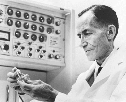
Photograph: Camillo Artom, M.D. working in lab circa 1960s (From the Subject File Collection)
Size: 5 cubic feet
Restrictions: The papers of Dr. Camillo Artom are open to qualified scholars after an interview with the Archives’ staff.
Preservation: Staples, rubber bands, and paper clips have been removed. Photos have been placed in Mylar sleeves and older documents interlaced with acid free paper. The entire collection has been placed in Hollinger boxes. Number of boxes: 13Provenance: The papers of Camillo Artom, M.D., Ph.D. were given to the Dorothy Carpenter Medical Center Archives, Coy C. Carpenter Library, Bowman Gray School of Medicine, on July 26, 1974 by Bianca Artom.
Biography:Dr. Camillo Artom, know as the "fat" chemist for his work with lipids, was born in Asti, Italy on June 6, 1893. After studying medicine at the University of Rome and the University of Padua, he received his M.D. from Padua in 1917. Artom served as a cadet sergeant and later a lieutenant in the Italian medical corps from 1916 to 1920. He was awarded the Italian and Romanian Crosses for his work terminating a typhus epidemic during his time in the service. Dr. Artom was also a skilled mountain climber, a talent much appreciated by the Alpine troops.
In 1923 he received his Ph.D. from the University of Messina and in 1926 a Ph.D. in Biochemistry from the University of Palermo. Although Artom earned a M.D., he only practiced medicine during the time he spent in the Corps. After the Great War or World War I, Artom took positions in the Universities of Messina and Palermo. During this ten-year period, he also conducted research at the Universities of Amsterdam and Frankfurt. According to an article in the Winston-Salem Journal and Sentinel from September 24, 1967, "In 1930, he became chairman of the department of biochemistry at the University of Cagliari, and in 1935, he took the same position at the University of Palermo in Sicily." He became a research fellow with the Rockefeller Foundation in Naples around the same time. At the time of this appointment, Dr. Camillo Artom was considered one of the foremost biochemists in Europe.
According to the same article in the Winston-Salem Journal, by the late 1930s Artom and other Jews in Italy were beginning to be aware of the negative feeling toward Jews in Europe. Artom described the situation: "By 1938, we were aware of the plight of the Jews in Germany. We did not expect the same trouble in Italy, for we Jews were so few." However, after Artom was denied permission to attend a conference of biochemists in Zurich, Switzerland, and dismissed from his position at the University of Palermo he realized the enormity of the situation, "I knew then that it was time we left Italy. There was nothing there for us anymore." Dr. Artom and his wife, Bianca, did not have an easy time leaving Italy. Working through the American consul in Naples, Artom sought to be included in the 1938 or 1939 quota of Italian immigrants to the United States. At the same time he worked relentlessly to secure a passport. Unfortunately, Artom would not be included in the quotas without proof of a job. Dr. Coy Carpenter, dean of Wake Forest Medical School secured that job. Dr. L. Emmett Holt of Johns Hopkins in Baltimore, Maryland recommended Dr. Artom to Dr. Carpenter. Carpenter offered Artom the position of chairman of the Biochemistry Department at Wake Forest Medical School. As the head of this one-man department, Artom was paid a meager salary of $200 month. Unfortunately, when Artom presented Dr. Carpenter’s cable offering him the position, the Italian consulate was furious. Dr. Artom was not allowed the leave because he was told his presence in the United States would provide unfair competition to the citizens of the United States. Fortunately, for Wake Forest Medical School, men of academic talent were exempted from the quota restrictions. Dr. Artom and his wife were allowed to move to the United States in 1939. Artom questioned their ability to live on such a low salary, but was assured it was possible in the small town of Wake Forest in North Carolina. Years later, despite offers from such prestigious universities as the Massachusetts Institute of Technology, Dr. Artom continued at Wake Forest. When asked why he remained in such a rural setting, Artom replied, "[t]his school helped me in a difficult time of my life. I felt an obligation to stay here." Dr. Artom was best known for his work in biochemistry, specifically lipid metabolism. Lipid metabolism deals with how the body absorbs fats, how the liver processes fats, and how fats collect in the walls of the arteries. Artom’s explanation of how fats are digested is still accepted today.Artom discovered that if the liver is not supplied with choline, a vitamin B-like substance, then the liver produces fatty tissues, a condition similar to cirrhosis. Working with rats and later humans, Artom and two other doctors, Cayer and Cornatzer, discovered that an injection of choline helped to reduce this condition. Dr. Artom also demonstrated that in the digestion of fats, mono- and diglycerides are formed. It is these forms of fats that are digested in the intestine.
During the 1960s, Artom used pigeons in his study on atherosclerosis. During this time, hardening of the arteries was considered the number one killer in the United States and Europe. For this reason, alone, Artom’s work was considered extremely important. Other landmark research by Artom included using radioactive isotopes. Artom used a radioactive isotope of phosphorus and injected it in a rat to trace the movement of phosphorus in the body. In the span of his professional life Dr. Artom published over 200 papers relating to his work and received numerous grants, even after age 70. His only hobby as professor of emeritus seemed to be reading the Journal of Biochemistry. According to Artom in 1967, "it is no longer possible to be a cultivated biochemist. The explosion of knowledge in biochemistry is so vast, so rapid, a man has time only to keep pace with his own field. It is so in my field which, is narrower than I like." In a nomination for the Albert and Mary Lasker Foundation’s 1966 Medical Research Award, Irving Carlyle writes,| "The nature of Dr. Artom’s contribution [to science] cannot be stated as a single discovery or technique. Rather, it is the comprehensive, wide-nature of his investigations that have made them significant advances all across the field of fat metabolism." |
In addition to professor and head of the department of Biochemistry at Wake Forest College School of Medicine from 1939 to 1942 and the professor and head of the Department of Biochemistry at Bowman Gray School of Medicine from 1941 to 1963, Dr. Artom was a member of various societies and held many offices. These include the:
Dr. Artom served as Emeritus Professor from 1963 until his death on February 3, 1970. Dr. Artom has been described as a faithful, humble, man totally lacking any "meanness" of spirit. His colleagues described him as intense but serene, a man that always worked diligently whether he was in the lab or in the classroom. In 1963 the graduating class of the Bowman Gray School of Medicine dedicated their yearbook, The Gray and White Matter to Dr. Artom, "an internationally known scientist (who) has engendered our awe and admiration."
Scope of the Collection: The papers of Camillo Artom, M.D., Ph.D. reflect, in part, the professional and personal life of one of Europe’s most prestigious biochemists from the time he and his wife left Italy until Dr. Artom’s death in 1970. There are documents in the collection that are dated before 1939, the date of Dr. Artom’s arrival in the United States, however, they are few.
The Artom Collection is primarily a collection of research notes and recorded data. However, there are teaching materials; evaluations of people and papers; papers presented at conferences and symposiums; correspondence and an address book; and, finally, items that recognize Dr. Artom’s achievements. These series comprise a partial collection that focuses on the professional life of Dr. Camillo Artom.
As one of Europe’s leading biochemists, Dr. Artom’s research with lipids and lipid metabolism was on the cutting edge. The main body of research notes have been arranged by subject according to Dr. Artom, while the remainder of the notes have been arranged artificially according to subject and alphabetical order. Other research materials include two boxes of bibliographic note cards, reprints related to Dr. Artom’s work, and reprints of articles that have been written by Dr. Artom or in collaboration with Dr. Artom. The collection also contains several unusual items. The items include a piece of rubber tubing with strings attached to the opposite ends, notes written on paper towels, and an address book. The address book is labeled "Rubrica" and contains the names and addresses of Artom’s friends and scientists.
The collection has been arranged into seven series:The papers in Dr. Artom’s collection have been arranged artificially with two exceptions: files 55 through 100, in boxes 5-9, and the note cards. These groups have been retained according to the arrangement by Dr. Artom. Also noted is the correspondence interspersed throughout the papers, in addition to the series labeled correspondence.
Series Description:
Series I. Curriculum Vitae, Bibliography, and Publications, Box 1, Files 1-5.
Series one consists of the curriculum vitae of Dr. Camillo Artom and rough draft of the curriculum vitae of Dr. Hugh Lofland. This series also consists of Dr. Artom's bibliography, revised and updated through the years, as well as a list of his publications. It should be noted that there are four bound volumes of Dr. Artom's writings. These volumes contain Xeroxed copies of reprints.
Series II. Research Notes, Box 1-13, Files 6-131.
The research notes have been artificially arranged according to subject. File numbers 6-54 have been artificially and then alphabetically arranged. The last three files that have been arranged artificially are the notebooks. These books contain data and research findings. They may have been used for class or in the lab.
Numbers 55-131 have been retained according to Artom’s arrangement. These notes contain the findings of Dr. Artom’s research on lipids and lipid metabolism. The files contain notes, graphs, charts, and papers reporting his results. The numbers at the end of each file label indicate the number of the file placed there by Dr. Artom. Interspersed throughout the notes and data is correspondence concerning the research in that particular file.
Within Dr. Artom’s arrangement there are three files of materials he collected concerning the changing nature of science. These files are labeled, "Science in a Changing World," "Science and Politics 1963-1964," "Serendipity-Science in a Changing World." Two other files not directly related to research under this series are labeled, "Federation proceedings."
Other research materials include two boxes of bibliographic note cards. Box 1-A, Dr. Artom’s label, has been arranged alphabetically according to the author. Also found in box 1-B were advertisements and postcards from the 1950s. Dr. Artom also collected reprints of other’s work. The final file under this series is an extensive collection of reprints of journal articles written by Artom or in collaboration with Artom.
Series III. Teaching Materials, Box 14, Files 132-138.
Series III reflects the teaching aspect of Dr. Artom’s career. These files contain experiments assigned to the students; lecture notes; a file on the History of Medicine Society; and two files labeled, "Advanced Topics in Biochemistry, 1966, and "Biochemistry Course, 1968,". There are also two theses within this series by Marjorie Swanson and Margaret Mitchell.
Series IV. Evaluations, Box 14, Files 139-141.
Series IV consists of correspondence inviting Dr. Artom to evaluate other doctors and scholarly works. The file also contains correspondence requesting that Dr. Artom evaluate faculty for promotion.
Series V. Conferences, Retreats, and Symposiums, Box 15, Files 142-146.
This series contains documents generated in response to the conferences, retreats, and symposiums attended by Dr. Artom. Such documents include papers, receipts, lists, and other items concerning travel arrangements.
Series VI. Correspondence and Address Book, Boxes 15-16, Files 147-153.
Although correspondence is interspersed throughout the collection, this series contains only personal and professional correspondence. The correspondence that is grouped according to subject concerns grants and experiments as well as the sale of back journals. The final item in this series is the address book, which contains the addresses of scientists, colleagues, and friends from around the world.
Series VII. Recognition, Box 16, Files 154-156.
This series documents, in part, the achievements of Dr. Artom. This series contains an encyclopedia entry written by Dr. Artom explaining lipids, a handwritten speech accepting the portrait dedicated to Dr. Artom, and a speech given to the Torch Club, a professional organization of distinguished members who are asked to share, on a monthly basis, their work to be commented on.
Inventory:
| Series I. Curriculum Vitae, Bibliography, and Publications, Box 1, Files 1-5 | ||
|
Box 1: |
||
|
File 1: Curriculum Vitae of Camillo Artom and Hugh Lofland, 1930-1950 File 2: Bibliography Rough Draft, 1914-1966 File 3: Artom publications, 1913-1969 File 4: Bibliography, 1945-1947 File 5: Revised Bibliography, 1969-1971 |
||
| Series II. Research Materials (Artificial Arrangement), Boxes 1-13, Files 6-131 | ||
| Box 1: | ||
| File 6: Choline [oversized, Box A]
File 7: Choline (DTMs) C14 Methyl labeled Dimethylaminoethanol [oversized, Box A] File 8: Choline phosphokinase File 9: Coconut Oil in human feeding File 10: Enzymes for Phospholipid Synthesis Experimental Results [oversized, Box A] File 11: Enzymes for Phospholipid Biosynthesis PC Diglyceride Transferase Summary (undated) File 12: Cytioyl Transferase Glyceride Transferase (undated) File 13: Paper by Raymond Reiser "The Intestinal Absorption of Triglycerides" File 14: Two Pathways (Fatty Liver Statistical Treatment) [oversized, Box A] File 15: Two Pathways for Lecithin Formation=B.B. Aeta Manuscript, 1950s File 16: Two Pathways – Manuscript 1967 slices File 17: Two Pathways Normal (Experimental Data Notes) [oversized, Box A] File 18: Drafts of paper—Seminar, January 1969 "Pathways for Lecithin" File 19: Lecithin Formation in Rat Tissues, 1964 File 20: Lecithin Formation in Rat Liver Slices (charts) [oversized, Box A] File 21: Two Pathways for Lecithin Formation, undated File 22: Two Pathways (Fatty Livers Statistical Treatment) [oversized, Box A] File 23: Enzymes for the Formation of Lecithins by Transmethylation the Livers of Developing Rats, 1969 |
||
| Box 2: | ||
| File 24: Reprints from others work on
Ethionine, 1950s
File 25: Manuscript – "Metabolism Phospholipid" File 26: Phospholipid Metabolism, 1953 File 27: Abstracts of proofs – Incorporation of Amino Acids Methyl C., 1965 File 28: Notes on lipid metabolism in Rat Liver Slices, 1960 [oversized, Box A] File 29: Future Experiments on lipid metabolism, 1960 [oversized, Box A] File 30: Metabolism of Phospholipid, undated File 31: Enzymes for Phospholipid, 1968 File 32: Phospholipid levels in liver disease, 1952 File 33: Paper "Ionizing, Radiation, Atherosclerosis, and Lipid Metabolism in Pigeons," undated File 34: Data used in Manuscript of Pigeon paper [oversized, Box A] File 35: Correspondence - "Ionizing, Radiation, Atherosclerosis, and Lipid Metabolism in Pigeons," 1960 File 36: Galley Proofs Var-Drafts Proofs of the paper, 1960 File 37: Enzymes for Lecithin Formation by Transmethylation in developing rats, final manuscript, undated File 38: Phosphatidye Choline by Transmethylation, undated File 39: Liver Data – Methionine Activity and Methyl Transferase File 40: TLC and paperchromatography Methyl Transferase, undated File 41: Identity of Methionine 4 Methyl Transferase in liver and lungs File 42: Methyl Transferase Data Graphs and Notes File 43: To Biophysical and Research communications, 1965 File 44: Determination of MME – Notes |
||
| Box 3: | ||
| File 45: Additions of DME and MME –
Notes, 1962
File 46: Preparation of C 14 Methyl DNA and other, 1950s File 47: Methylations in Metabolism, 1950s File 48: Charts and Graphs, 1950s File 49: Camera Ready Prints of Charts File 50: Tables and drafts, "Incorporation of the Carbons of L-[14 C] -Methionine Into the Lipids of Rat Intestinal Mucosa." |
||
| Box 4: | ||
| Files 51-54 Notebooks, 1030s, Files 43-45 in Box 4 Artom's Arrangement | ||
| Box 5: | ||
| File 55: Serine phospholipid (complete
manuscript) 1, 1964
File 56: Serine incorporation into phospholipid 2, 1962 File 57: Two pathways – liver slices experimental results 3, undated File 58: Two pathways of lecithin formation – future experiments 4, undated File 59: Science in a changing world 6, 1963 File 60: Science and politics, 1963 and 1964 File 61: Serendipity – Science in a Changing World 8, 1964 File 62: Serine incorporation into lipids preliminary report 9 File 63: Serine incorporation into phospholipid 10, 1962 File 64: EA and Serine Incorporation into phospholipid and Experimental Results 11 [oversized in Box A] File 65: MME phospholipid preliminary reports 12, 1960 File 66: MME phospholipid B and B research communications 13, 1964 File 67: MME phospholipid 14, 1961 File 68: MME phospholipid, literature, and methods 15, 1963 File 69: MME phospholipid-summaries of experiment results 16, 1963 |
||
| Box 6: | ||
| File 70: Orotic acid ethionine fatty
livers, 1963
File 71: EA and PEA 18 [oversized in Box A] File 72: DME = experimental data (summary) 19, undated File 73: DME, 1961 File 74: Choline determination [notes on paper towels] 21, 1966 File 75: Col-stimulation of phospholipid formation 22, 1963 File 76: Atp Dtm, 23 File 77: Aminoeaeyl-lipids experimental results 24, 1959 File 78: Experiments in progress (April 1, 1960) 25 [oversized Box A] File 79: DME Reprints 28, 1960s File 80: Reprints for current investigations, 1960s File 81: Enzymes of lecithin formation seminar, January 1968 File 82: Experiment Results – enzymes of lecithin information CCl14 ethionine effects 31, 1967 File 83: Current Experiments 1967 32 File 84: Not labeled [reprints], 1950s and 1960s File 85: Seinis N.D., 34 1961 File 86: Peptidolipids 35 1966 File 87: Outside of file correspondence about methione Methyl-C-14, 1968 |
||
| Box 7: | ||
| File 88: Two pathways of lecithin
formation results and Calculations on permeability 36, undated
File 89: PC-Glyceride transferase choline phosphokinase reprints 37, 1950s and 1960s File 90: Aminoacyl lipids 1960 experiments 38 File 91: Two pathways liver slices 1965 statistical treatment of data 39 File 92: Methionine activating enzymes 40 1965 File 93: Liver enzymes (relative values and statistical evaluations) 41, undated File 94: Lung lecithin 42, 1960s File 95: Enzymes for phospholipid biosynthesis choline Kinase cytidyl transferase 43 File 96: Two pathways for lecithin formation seminar (v. of n.d.) 44 File 97: Federation proceedings, 27 (2), 457 (1968) File 98: Federation proceedings, 28, 845, (1967) |
||
| Box 8: | ||
File 99: Note cards Box 1-A, 1913-1970
|
||
| Box 9: | ||
File 100: Box 1-B: Note cards missing a
label, possibly labeled under B
Notecards Bibliography, B-W, arranged alphabetically by author's last name Reprints and other works collected by Artom |
||
| Box 10: | ||
| File 101: 1946
File 102: 1950-1951 File 103: 1952-1953 File 104: 1954-1955 File 105: 1956-1957 File 106: 1958 File 107: 1959 File 108: 1960 Artom Reprints |
||
| Box 11: | ||
| File 109: undated reprints
File 110: Paper written in 1918 File 111: 1910-1915 File 112: 1916-1920 File 113: 1921-1923 File 114: 1924-1925 File 115: 1926-1927 File 116: 1928-1929 |
||
| Box 12: | ||
| File 117: 1930-1932
File 118: 1933, 1935, and 1936 File 119: 1937 File 120: 1938 File 121: 1940, 1941, and 1942 File 122: 1943 and 1944 |
||
| Box 13: | ||
| File 123: 1945 and 1946
File 124: 1947, 1948, and 1949 File 125: 1950 and 1951 File 126: 1954 and 1955 File 127: 1954 and 1955 File 128: 1956 and 1957 File 129: 1958 and 1959 File 130: 1960, 1961, and 1962 File 131: 1964, 1965, 1966, 1967, 1968, 1969 |
||
| Series III. Teaching Materials, Box 14, Files 132-138 | ||
| Box 14: | ||
| File 132: Student Assignments (lipid
experiments)
File 133: Lecture Notes [oversized Box B] File 134: Thesis by Marietta Crowder, 1948 File 135: Thesis by Margaret Mitchell, 1948 File 136: Advanced Biochemistry Class 1966 File 137: Biochemistry Course 1968 File 138: History of Medicine Society, 1960s |
||
| Series IV. Evaluations, Box 14, Files 139-141 | |
| File 139: Requests to evaluate people for
awards, 1940s and 1950s
File 140: Requests to evaluate people for papers, 1950s and 1960s File 141: Faculty Promotions, 1950s and 1960s |
|
| Series V. Conferences, Retreats, and Symposiums, Box 15, Files 142-146 | ||
| Box 15: | ||
| File 142: Paper given in New Orleans,
1940
File 143: Lipid Symposium, 1950 File 144: Faculty Retreat, 1958 File 145: Lipotropic Symposium, 1958 File 146: Deuel Conference on Lipids, 1962 |
||
| Series VI. Correspondence and Address Book, Box 15-16, Files 147-153 | ||
| Box 15 | ||
| File 147: Personal and Professional, some
in Italian, 1935-1966
File 148: Personal and Professional, some in Italian, 1960 |
||
| Box 16: | ||
| File 149: Speech and visit by Dr. Severo
Ochoa and Dr. William Rose, 1962
File 150: Grants and Experiments, 1950s and 1960s File 151: Sale of Back Journals, 1969 File 152: Address Book File 153: Miscellaneous items interspersed with correspondence |
||
| Series VII. Recognition, Box 16, Files 154-156 | ||
| Box 16: | ||
| File 154: Encyclopedia Entry, 1949, [also
oversized, Box B]
File 155: Portrait Dedication, 1969 [oversized, Box B] File 156: Torch Club, 1960s [oversized, Box B] Addendum: May 1994 Box of Research Notebooks generated by Dr. Artom |
||
Series:
Back to the top.
Back to Archive Collections.
Introduction: While this finding aid encompasses the entire personal collection of Dr. McHenry, it focuses on the slides and photographs in the collection. A description of each series in the large collection is included, as well as a detailed inventory of the slides and photographs interspersed throughout the collection.
Size: 207 cubic feet
Restrictions: Most of the papers of the Lawrence Chester McHenry, Jr., M.D. Collection are open to qualified scholars. Any questions concerning access should be directed to The Dorothy Carpenter Medical Archives Archivist. Materials of an extremely personal nature within this collection are restricted to outside use. See below series descriptions for more information.
Preservation: Materials have been placed in alkaline folders in alkaline boxes. Slides and photographs have been placed in Mylar sleeves.
Number of boxes: 64
Provenance: The papers of Lawrence Chester McHenry, Jr., M.D. were donated to The Dorothy Carpenter Medical Archives after Dr. McHenry's death on February 22, 1985.
Biography: Lawrence Chester McHenry, Jr. was born March 20, 1929 in Oklahoma City, Oklahoma. He was the second child of Dr. Lawrence Chester McHenry, Sr. and Hilda (Pentland) McHenry. He grew up in Oklahoma City and received his earliest education there. In 1944, he went to the New Mexico Military Institute, where he received his high school diploma in 1947. In 1951 he received a B.A. from Pomona College in Claremont, California. Returning to Oklahoma City, he entered medical school at the University of Oklahoma in 1951 and graduated in 1955 with a M.D. degree. From Oklahoma he went to Boston, Massachusetts for internship and residency.
While in Boston, 1955-1960, he studied at Harvard University; worked at the Massachusetts General Hospital, Boston City Hospital, and the New England Center Hospital; and married Anne Marie Riedl. From this marriage three children were born: Susan, 1958; Barbara, 1960; and Robert, 1964. In 1968 he and Anne Marie Riedl divorced and the following year, 1969, he married Kathryn Olson. During his medical school, internship, and residency years, Dr. McHenry developed an interest in two areas that remained with him his entire life: the history of medicine and the life of Samuel Johnson. After serving at Walter Reed Army Hospital from 1960-1964 as a captain in the U.S. Army Reserve, Dr. McHenry’s career centered around research into cerebral blood flow measurement, stroke, and cerebrovascular diseases.Biographical Notes
Born March 20 in Oklahoma City, Oklahoma (1929)
Graduated from high school, New Mexico Military Institute (1947)
B.A., Pomona College, Claremont, California (1947-1951)
M.D., School of Medicine, University of Oklahoma, Oklahoma City, Oklahoma(1951-1955)
Summer Study, Deutschkurse fur Auslander, Akademischen Auslandstelle, University of Munich, Munich, Federal Republic of Germany (1953)
Licensed to practice medicine, State of Oklahoma (1955)
Membership, American Association History of Medicine (1955)
Medical Intern, Boston City Hospital, Boston, Massachusetts (1955-1956)
Assistant Resident in Medicine, Boston City Hospital, Boston, Massachusetts (1955-1957)
American Academy of Neurology, Fellow (1957)
Married Anne Marie Riedl (1957)
Assistant Resident in Neurology, New England Center Hospital (1957-1958)
Fellow in Neurology, Tufts University School of Medicine, Boston, Massachusetts (1957-1958)
Birth of first child, Susan P. McHenry (October 3, 1958)
Resident in Neuropathology, Boston City Hospital, Boston, Massachusetts (1958-1959)
Fellow in Neurology, Harvard Medical School, Cambridge, Massachusetts (1958-1959)
Induction into the Johnson Society, Lichfield, England: Life Member (1959)
Resident in Neurology, New England Center Hospital, Boston, Massachusetts (1959-1960)
Fellow in Neurology, Tufts University School of Medicine, Boston, Massachusetts (1959-1960)
Birth of second child, Barbara A. McHenry (May 16, 1960)
Summer Study, Visiting Fellow, History of Medicine Department, Yale University Graduate School, New Haven, Connecticut (1960)
Captain, MC USAR, Department of Neurophysiology, Division of Neuropsychiatry, Walter Reed Army Institute of Research, Washington, D.C. (1960-1964)
Clinic Supervisor, Neurology Service, Walter Reed General Hospital, Walter Reed Army Medical Center, Washington, D.C. (1961-1964)
Clinical Instructor in Neurology, George Washington University, Washington, D.C (1961-1964)
Certification, American Board of Psychiatry and Neurology (1963)
Induction into Membership in the Johnson Society of London (1963)
Membership, Philadelphia Neurological Society (1964)
Licensed to Practice Medicine - State of Pennsylvania (1964)
Birth of third child, Robert L. McHenry ( June 7, 1964)
Assistant Attending Physician, Philadelphia General Hospital, Philadelphia, Pennsylvania (1964-1965)
Chairman, The Johnsonians (1965)
Major, MC USAR (1965-1968)
Assistant Professor, Jefferson Medical College, Philadelphia, Pennsylvania (1965-1968)
Chief of Service (Jefferson), Philadelphia General Hospital, Philadelphia, Pennsylvania (1965-1971)
Neurologist, Jefferson Medical College Hospital, Philadelphia, Pennsylvania (1965-1971)
Fellow, Royal Society of Medicine, London, England (1966)
Fellow, American Academy of Neurology (1966)
Secretary, Philadelphia Neurological Society (1966-1970)
Director, Stroke Research Center, Philadelphia General Hospital, Philadelphia, Pennsylvania (1966-1972)
John and Mary R. Markle Scholar in Academic Medicine (1967-1972)
Divorced from Anne Marie Riedl (1968)
Marriage to Kathryn Olson McHenry (1969)
Associate Professor, Jefferson Medical College, Philadelphia, Pennsylvania (1969-1971)
Member, American Neurological Association (1969)
Visiting Professor of Neurology, University of Alabama, Birmingham, Alabama (1970)
President, Philadelphia Neurological Society (1971)
Associate Professor of Neurology, University of Pennsylvania, Philadelphia, Pennsylvania (1971-1972)
Senior Attending Physician, Philadelphia General Hospital, Philadelphia, Pennsylvania (1971-1972)
Staff, Department of Neurology, Hospital of the University of Pennsylvania, Philadelphia, Pennsylvania (1971-1972)
Licensed to Practice medicine - State of North Carolina (1972)
Professor of Neurology, Bowman Gray School of Medicine, Winston-Salem, North Carolina (1972-1985)
Who’s Who in the East (1972-1975)
Chairman, The Johnsonians (1973)
Co-Chairman and Editor, The Sixth International Symposium on Cerebral Blood Flow, Philadelphia, Pennsylvania (1973)
Who’s Who in the South and Southeast (1976-1984)
Membership, American Osler Society (1978)
Visiting Professor of Neurology, The Medical College of Pennsylvania, Philadelphia, Pennsylvania (1979)
Membership, Forsyth-Stokes-Davie County Medical Society (1981)
Membership, North Carolina Medical Society (1981)
American Neurological Association - Historian and Archivist (1981-1985)
Member, American Medical Association (1982)
Member, Editorial Advisory Board, The Classics of Medicine Library1982
Board of Trustees, Unitarian-Universalists Fellowship of Winston-Salem, North Carolina (1982-1983)
American Academy of Neurology, Chairman, Section on the History, of Neurology (1982-1984)
North Carolina Medical Society, Committee on Physicians Health and Effectiveness (1982-1985)
Board of Governors, North Carolina Jewish Home, Clemmons, North Carolina (1982-1985)
Chairman, Editorial Advisory Board, The Classics of Neurology and Neurosurgery Library (1983)
American Association History of Medicine, Chairman, Osler Medal Committee (1983)
Membership, Johnson Society of the Central Region, U.S.A. (1983)
World Federation of Neurology, History of Neurosciences Research Group, American Secretary (1983)
Member, College of Physicians of Philadelphia (1983)
Vice-President, American Osler Society (1984)
Member, Advisory Council on Addictive Diseases, Charter-Mandala Center, Winston-Salem, North Carolina (1984-1985)
Died February 22 in Winston-Salem, North Carolina (1985)
Scope of the Collection: The papers of Lawrence Chester McHenry, Jr., M.D. cover his life, education, and work from 1936 to February 1985. Included in the collection are personal and family papers; college, medical school, internship, and residency records; military orders and credentials; medical research, scientific investigation, and Cerebral Blood Flow Studies; diaries, documents, and discussions related to Alcoholics Anonymous; notes and manuscripts dealing with the history of medicine, the history of neurology, and the medical aspects of the life of the Eighteenth Century English writer, Samuel Johnson; reprints of articles, manuscripts, correspondence, patient studies, and photographs.
The collection provides both an overview of and specific information on Dr. McHenry’s personal life, medical career, scientific research, and scholarly writings. From the papers pertaining to Dr. McHenry’s personal life, his growth and development as a medical doctor, scholar, writer, and historian is seen. His military records document his career as an officer in the United State Army. While his medical research information provides insight into neurological clinical research and experimentation, the papers that deal with the history of medicine contain Dr. McHenry’s insights as a writer, scholar, and scientist.
Dr. McHenry’s professional associations show his involvement not only with the scientific aspects of medicine but also with the historical features of medical endeavor. His professional relationships are catalogued within committee reports, correspondence, and lecture notes. In its totality, this collection of papers illustrates the life, work, and interests of a man who was multi-dimensional.
The collection is arranged in eight series:
The arrangement of the material in all eight series adheres as closely as possible to the order in which the papers were received from Dr. McHenry in August 1984 and from his office after his death in February 1985. In many instances folders have been kept intact and original folder headings have been retained.
Summary of Inventory
Series I. Personal and Family Papers, 1-70.
Series II. Scrapbooks, 71v-91v.
Series III. Correspondence, 92-454.
Series IV. Professional Papers: Reprints, 455-701V.
Series V. Professional Papers: Research, 702-748v.
Series VI. Professional papers: Employment, 749-767.
Series VII. History of Neurology, 768-929
1. American Academy of Neurology (AAN)
2. American Association of the History of Medicine (AAHM)
3. American Neurological Association (ANA)
4. Philadelphia Neurological Society (PNS)
5. World Federation of Neurology (WFN)
6. Gryphon Editions, Inc.
a. Classics in Medicine
b. Classics in Neurology
History of Stroke. 813-822.
American Neurology. 823-859.
Neurology and Art. 860-867.
1. Charles K. Mills, 1845-1931.
2. William Osler, 1849-1919.
3. S. Weir Mitchell, 1829-1914.
4. William Alexander Hammond, 1828-1900
5. Francis X. Dercum, 1856-1931.
6. Eadweard Muybridge, 1830-1904.
7. Origins of American Neurology and Hospitals
Garrison’s History of Neurology. 868-910.
Philadelphia Orthopedic Hospital and Infirmary for Nervous Diseases. 911-929.
Series IX. Samuel Johnson, 930-1007 (Kept with Dr. McHenry’s original arrangement).
Series Description/Inventory
Series I. Personal and Family Papers, Files 1-79
The series presents a detailed overview of Dr. Lawrence Chester McHenry’s life, family, and ancestors. These papers chronicle his growth and development as a student, doctor, researcher, and scholar, as well as recording his experiences as a husband and father. Both the public documentation and the private accounts of his life are included in this collection.
The large amount of materials present has necessitated this series being divided into four sub-series: McHenry Family History; Lawrence Chester McHenry, Jr., Education; Lawrence Chester McHenry, Jr., Military and Lawrence Chester McHenry, Jr., Personal. Though some refoldering has been necessary, the order and arrangement of the papers are primarily as Dr. McHenry intended.
Subseries A, McHenry Family History, 1-13, contains papers about the genealogical aspects of several branches of the McHenry Family. Also included are early records (1817), family tree and coat of arms, correspondence, and family information. Specific materials on Dr. Lawrence McHenry Jr.'s grandfather and father and mother are found in these folders.
The papers found in subseries B, Lawrence Chester McHenry, Jr.: Education, 13-32, are grouped into six areas: primary and secondary schools, college, medical school, internship and residency, Markle Scholarship Program, and Continuing Medical Education. Education was very important to Dr. McHenry and he kept many of the papers associated with his varied experiences. Thus, the reader is presented with a full picture of his educational background. Contained in these folders are report cards, transcripts, certificates, diplomas, reports of social activities, early writings, schedules, lecture notes, patient information, correspondence, and forms.
Lawrence Chester McHenry, Jr.: Military, subseries C, 33-39, includes both the formal and informal records of Dr. McHenry's military experiences. These folders contain his induction notice, ROTC history, correspondence, health records, general orders, duty reports from the Walter Reed Army Institute of Research, and certificate of honorable discharge.
The last subseries in Series I, is subseries D, Lawrence Chester McHenry, Jr.: Personal. Materials in this subseries are restricted and are not available for public use.
Because of the personal nature of some of the materials contained in this collection, certain materials interspersed within the subseries are restricted and the entire subseries, Lawrence Chester McHenry, Jr., Personal is restricted. An interview with the Archivist is required before viewing any materials in this collection.
Series II. Scrapbooks, 71v.-91v.
This collection of Dr. McHenry’s personal and professional scrapbooks is divided into four subseries that cover the following areas: personal and family events, research, the History of Neurology, and Samuel Johnson.
These scrapbooks are the result of Dr. McHenry’s collecting, arranging, and maintaining the tangible items significant to the various segments of his life and work. All of the scrapbooks are as Dr. McHenry arranged them and include only the materials he felt were important. Their order and content are exactly as Dr. McHenry prepared them.
Subseries A, Personal Scrapbooks, 71v.-75v., is comprised of various memorabilia that reflect Dr. McHenry’s life from the 1930’s until 1982. These scrapbooks are in chronological order and contain; family photographs, school certificates (Oklahoma City and NMMI), regulations, handbooks, science fair experiments, and publications, correspondence (Moorman, Sigerist, medical school materials, maps, SITA travel booklets, and research notes associated with Walter Reed Army Institute of Research (WRAIR)), programs from meetings of the American Association of Neurologists, teaching and Continuing Education programs from George Washington University and Walter Reed, Markle Scholarship Award information (1967), annual reports, memos, name tags, and letters from Stroke Conferences, CBF Symposiums, and the Philadelphia Neurological Society, information from Appalachian Hall, staff directories for the Bowman Gray School of Medicine, and letters concerning the McHenry Collection in the Medical School Library.
Subseries B, Research, 76v.-83v. is, for the most part, the files of Cerebral Blood Flow and Stroke Research the Dr. McHenry conducted at Walter Reed and in Philadelphia at the Stroke Research Center. These scrapbooks illustrate Dr. McHenry’s presentation, arrangement, and perception of his research. This order, selection, and grouping is as Dr. McHenry arranged it thus, this subseries is dependent upon Dr. McHenry's interpretation of his research data. Included in the contents of these scrapbooks are: grant proposals, photographs, correspondence, reprints, budgets, minutes of planning meetings, equipment lists, diagrams, graphs, tables, and charts relating to the Krpton-85 method, formulas, patient studies, reports on the CBF labs of Ingvar (Lund), Lassen (Copenhagen), and Veall (London), and slides. These almost 400 slides illustrate the techniques, methods, procedures, and materials Dr. McHenry used with Stroke and CBF research from 1960-1972.
Slides in the Subseries include:
Scrapbook,
|
79v. "Slides, CBF, Stroke" |
||
|
Pages 1 and 2: |
||
| Slides 1 -35: Slides used for presentation: "The Stroke Syndrome:Pathogenesis" | ||
Page 3: |
||
|
Slides 36-49: Second copy of some of the slides used for Presentation: "The Stroke Syndrome: Pathogenesis" Slide 50: Fig. 26 Fruit and Vegetable peeler (drawing) Slide 51: Spoon (drawing) Slide 52: Fig. 30 The Uses of Suction Cups (drawing) |
||
|
Page 4: |
||
|
Slide 53: Posterior Cerebral Artery Occlusion Slide 54: Basilar Artery Thrombosis Slide 55: Cerebral Embolism Slide 56: Illustration of brain Slide 57: Temporal Lobe Vascular Malformation Slide 58: Illustration of brain Slide 59: Sagittal Sinus Thrombosis Slide 60: Outline of Non-Vascular Causes of the Stroke Syndrome Slides 61-62: Various illustrations of brain Slide 63: Parasagittal Memingioma Slides 64-65: Various illustrations of brain Slide 66: Diagram of brains Slides 67-70: Various drawings of brain and cerebral artery |
||
|
Page 5: |
||
|
Slide 71: Chart: Onset and Course of Neurological Disease Slide 72: Diagram: Blood Supply of Brain-Neck Vessels Slide 73: Diagram: Normal Cerebral Blood Supply - Circle of Willis Slide 74: Diagram: Body Supply of Brain Slide 75: Diagram: Blood Supply to Deep Structures of Cerebral Hemispheres Slide 76: Diagram: Functional Anatomy of the Cerebral Hemispheres Slide 77: Outline: Major Factors Influencing Cerebral Blood Flow Slide 78: Outline: Stroke Syndrome in Infancy and Childhood Slide 79: Outline: Stroke Syndrome in Adolescence and Young Adulthood Slide 80: Outline: Stroke Syndrome in Middle and Old Age Slide 81: Chart: Stroke Syndrome in Adults Slide 82: Chart: Clinical Syndromes of Cerebrovascular Aherosclerosis Slide 83: Chart: Carotid Arterial System - Transient Ischemic Attacks Slide 84: Diagram: Small Infarct in Basal Ganglia Slide 85: Diagram: Large Infarct in Basal Ganglia or Internal Capsule Slide 86: Diagram: Middle Cerebral Artery Occlusion Slide 87: Diagram: Anterior Cerebral Artery Occlusion Slide 88: Chart: Extracranial Carotid Artery Narrowing or Occlusion Slide 89: Chart: Prognosis in Completed Strokes |
||
|
Page 6: |
||
|
Slide 90: Table 4: Hemodynamic Causes of Diffuse Cerebral Ischemia Slide 91: Table 1: Sites of Arterial Stenosis ad Occlusion in Cerebrovascular Ischemia Slide 92: Outline: Transient Ischemic Attacks Slide 93: Outline: Transient Ischemic Attacks, Differential Diagnosis Slide 94: Outline: Transient Ischemic Attacks, Underlying Pathophysiology - II Slide 95: Table 2: Incidence and cause of Mortality Among TIA Patients Slide 96: Outline: Transient Ischemic Attacks, Medical Treatments, Dipridmole Slide 97: Outline: Transient Ischemic Attacks, Medical Treatment, Aspirin Slide 98: Table 5: Results of 1,546 Carotid Operations Slide 99: Outline: "Not TIA" Slide 100: Outline: Transient Ischemic Attacks, Precipitating Factors Slide 101: Table 2: Classification of 120 Patients with Migrainous Accompainments Slide 102: Outline: Cerebral Embolism Slide 103: Table 3: Vertebral - basilar TIA: Presenting Symptoms in 54 Patients Slide 104: Outline: Transient Ischemic Attacks, Subclavian Steal Syndrome - SSS Slide 105: Outline: Transient Ischemic Attacks, Transient Global Amnesia - TGA Slide 106: Chart: Years after first TIA Slide 107: Table 2: Carotid Artery TIA: Presenting Symptoms In 133 Patients Slide 108: Outline: Transient Ischemic Attacks Slide 109: Chart: Years of Follow-Up |
||
|
Page 7: |
||
|
Slide 110: List: Transient Ischemic Attacks - Characteristics Slide 111: List: Transient Ischemic Attacks, Carotid Artery System Slide 112: List: Clinical Characteristics of Transient Ischemic Attacks Slide 113: Outline: Gross Classification of Cerebrovascular Disease Slide 114: List: Transient Ischemic Attacks, Vertebral-Basilar System Slide 115: List: Cerebral Embolism II Slide 116: List: Precipitating Factors of Transient Ischemic Attacks Slide 117: List: Cerebral Embolism I Slide 118: List: Transient Ischemic Attacks, Surgical Treatment II Slide 119: Outline: Transient Ischemic Attacks, Evaluation of Patient I Slide 120: List Transient Ischemic Attacks, Medical Therapy Slide 121: Outline: Transient Ischemic Attacks, Incidence and Stroke Relationships Slide 122: Outline: Hemodynamic Factors in Transient Ischemic Attacks Slide 123: Outline: Transient Ischemic Attacks, Anticoagulant Therapy II Slide 124: Outline: Transient Ischemic Attacks, Medical Treatment Anticoagulant Therapy I Slide 125: Table 13: Clinical Outcome Six Months Following Randomization by Treatment Group and History of Previous TIAS Slide 126: Table 3: Main Criteria For the Diagnosis of Late-Life Migrainous Accompaniments Slide 127: Table 1: Infarction Among Untreated TIA Patients Slide 128: Transient Ischemic Attacks, Surgical Treatment I Slide 129: Table 2: Anticoagulant Therapy and Transient Ischemic Attacks` |
||
Page 8: |
||
| Slides 130-148: Various tables, charts and images from the Cerebral Blood Flow Lab | ||
|
Page 9: |
||
|
Slides 149-152: Various outlines concerning Regulation of Cerebral Circulation Slide 153: Outline: Cerebral Metabolism Slide 154: List: Acute Stroke and Vasopressor Agents Slide 155: Table: Effects of Drugs on Cerebral Circulation in Cerebrovascular Disease Slide 156: Summary: Regulation of Cerebral Circulation Slide 157: Table: Cerebral Blood Flow Values Slides 158-160: Various charts from the Cerebral Blood Flow Lab Slides 161-162: Unidentified images |
||
Page 10: |
||
|
Slides 163-174: Various unidentified charts, drawings and images Slide 175: Summary: Methods of Cerebral Blood Flow Measurement Slide 176: Outline: Additional Methods of Cerebral Blood Flow Measurement Slide 177: Unidentified photograph Slide 178: Chart of brain activity Slide 179: Figure 1: Regional Cerebral Blood Flow Slide 180: Table 4: Regional Cerebral Blood Flow Slide 181: Figure 1: Regional Cerebral Blood Flow Slide 182: Table 4: Regional Cerebral Blood Flow |
||
Page 11: |
||
|
Slides 183-194: Various unidentified charts, drawings and images Slide 195: Image labeled, "Resting, Hyperfrontality" Slide 196: Image labeled, "Listen to spoken word" Slide 197: Image labeled, "Speaking; mouth tongue area" Slide 198: Image labeled, "Reading aloud" Slide 199: Image labeled, "Voluntary movement, hand-finger" Slide 200: Image labeled, "Visual perception sensory" Slide 201: Image labeled, "Move mouth, counting" Slide 202: Image labeled, "Reading silently" |
||
| Page 12: (Page of slides is labeled, "Kety-Schmidt CBF Values Method") | ||
|
Slide 203: Chart: Cerebral Blood Flow Studies Before and During Hyperventilation Slide 204: Table 2: Blood Flow and Oxygen Demands on Several Vital Organs Slide 205: Cerebral Metabolic Rate (CMRO2) Slide 206: Cerebral Metabolic Rate (CMRO2) Slide 207: Cerebral Vascular Resistance (CVR) Slide 208: Chart: Cerebral Blood Flow-Krypton Desaturation Method Slide 209: Chart: minutes of inhalation Slide 210: Cerebral Blood Flow Slide 211: Unidentified Chart Slide 212: Unidentified Chart Slide 213: Unidentified Drawing Slide 214: Chart: Cerebral Blood Flow Slide 215: Equations dealing with cerebral blood flow Slide 216: Chart: Cerebral blood flow - Krypton Desaturation Method Slide 217: Unidentified Chart Slides 218-219: Unidentified photographs Slide 220: Unidentified Charts Slide 221: Unidentified photographs |
|
|
Page 13: |
||
|
Slides 222-225: Various unidentified photographs and drawings Slide 226: Chart and image labeled, "Dementia, gray weight" Slide 227: Chart and image labeled, "Serial Study, MCA brocel. WB, Serial study, aphesia" Slide 228: Chart and image labeled, "Hemorrhage, Intracerebral hem." Slide 229: Drawing labeled, "Obristxe inhalation apparatus" Slide 230: Unidentified scan Slide 231: Diagram labeled, "rCBF method, McHenry PGH" Slide 232: Unidentified scan Slide 233: Chart: Fitted Head Curve Slide 234: Chart: Respired Air Curve Slide 235: Chart: Inhalation Slide 236: Table: Test-Retest Results of Xenon Inhalation Slide 237: Table: Normal Values For the Xenon Inhalation Cerebral Blood Flow Method of Obrist Slide 238: Table 4: Normal rCBF Values obtained by several authors by the Xenon-133 Inhalation method Slide 239: Table 2 and 3: Test-Retest Results of rCBF Measurements by the Xenon Inhalation method in 10 Patients and Results from Discriminate Analysis of 15 Normal Subjects and 10 Patients |
||
|
Page 14: |
||
|
Slides 240-258: Unidentified scans and photographs from Bowman Gray School of Medicine |
||
|
Page 15: |
||
|
Slide 259: Chart: Effects of CO2 Inhalation and of induced Hypertension on Regional Cerebral Blood Flow Slide 260: Graph: Effects of CO2 Inhalation and of induced Hypertension on Cerebral Blood Flow and Cardiac Factor Group I Slide 261: Charts: Effects of CO2 Inhalation and of Induced Hypertension on Cerebral Blood Flow and Cardiac Factor Groups I and II Slide 262: Graphs: Effects of CO2 Inhalation and of Induced Hypertension on Cerebral Blood Flow and Cardiac Factor Groups I and II Slide 263: Same as bottom of slide 261 Slide 264: Same as bottom half of slide 262 Slide 265: Illustration: CO2 responsive, intracerbral steal Slide 266: Illustration: CBF with Ischemia Slide 267: Labeled: "Xenon Clearance Curve" Slide 268: Figures I and II: Regional Cerebral Blood Flow Before and During CO2 Inhalation in Focal Vascular Disease I and II Slide 269: Figures I and II: Regional Cerebral Blood Flow Before And During CO2 Inhalation in Focal Vascular Disease I and II Slide 270: Graph: Effects of CO2 Inhalation on Regional Cerebral Circulation in Stroke Slide 271: Table: Effect CO2 Inhalation on Regional Cerebral Circulation in Stroke Patients with Focal Abnormalities Slide 272: Graph labeled, "Summary CO2 CBF focus, non-focus" Slide 273: Drawing: Effects of PaCO2 on Experimental Cerebral Ischemia Slide 274: Drawing: Intracerebral steal Slide 275: Graph labeled, "Autoregulation" Slide 276: Graphs: Cerebral Blood Flow, Metabolism and Cardiac Function before and during CO2 Inhalation in Normotensive Stroke Patients |
||
|
Page 16: |
||
|
Slide 277: Chart: Regional Blood Flow in Patients with Cerebral Infarction - Before and After Papaverine Slide 278: Table II: Regional Blood Flow - Before and After Papaverine Diffuse Cerebrovascular Disease Slide 279: Table: Papaverine Effect on Regional Cerebral Circulation in Stroke Patients with Focal Abnormalities Slide 280: Summary: Papaverine Effect on Regional Cerebral Circulation in Stroke Patients with Focal Abnormalities Slide 281: Scan labeled, "ACA occlusion" Slide 282: Graphs labeled, "CO2 rCBF PGHBH" Slide 283: Graphs: Effects of CO2 Inhalation and Hexobendine on Cerebral Blood Flow, Metabolism and Cardiac Function Slide 284: Charts: Effects of CO2 Inhalation on Regional Cerebral Blood Flow Slide 285: Chart: Effects of Hexobendine on Regional Cerebral Blood Flow Slide 286: Diagram not labeled Slide 287: Unlabeled diagram Slide 288: Scan and chart labeled, "Hexobenine MC Co2 rCBF" Slide 289: Figure I: Regional Cerebral Blood Flow Before and During CO2 Inhalation in Focal Vascular Disease I Slide 290: Figure II: Regional Cerebral Blood Flow Before and During CO2 Inhalation in Focal Vascular Disease II Slide 291: Charts: Effects of CO2 Inhalation and of induced Hypertension on Regional Cerebral Blood Flow Slide 292: Table: Hydergine Effect on Cerebral Circulation in Patients with Cerebrovascular Disease Slide 293: Chart: Hydergine Effect on Cerebral Circulation in Cerebrovascular Disease Slide 294: Unlabeled diagram Slide 295: Table: Effects of Drugs on Cerebral Circulation In Cerebrovascular Disease |
||
|
Page 17: |
||
|
Slide 296: Page from computer printout, rCBF Slide 297: List: Regional Cerebral Blood Flow Values: Criteria for significant change between repeat rCBF Measurements Slide 298: List: Regional Cerebral Blood Flow Values: Criteria for Determination of Abnormality Slide 299: Graphs: Serial study and comparative study Slide 300: Unidentified diagram Slide 301: Chart labeled: "Tissue perfusion pressure" Slide 302: Unlabeled drawing Slide 303: Unlabeled diagram Slide 304: Table: Local Cerebral Blood Flow in the Cat Slide 305: Unidentified images Slide 306: Figure 1: Normal Dilution Courses Slide 307: Unidentified diagram |
||
|
Page 18: |
||
|
Slide 308: Drawing: Probe Positions Slide 309: Charts: Flow in Rapid Clearing Compartment Slide 310: Charts: Flow in Rapid Clearing Compartment, Ischemic Episode - No Residual Slide 311: Charts: Flow in Rapid Clearing Compartment, Carotid Disease - Indefinite Symptoms Slide 312: Charts: Flow in Rapid Clearing Portion Slide 313: Table: Patient Group Slide 314: Table: Patients with Ischemic Infarction and Minimal Residual Deficit Slide 315: Table: Flow in Rapid Clearing Compartment in CC/100 GM/MIN Slide 316: Table: Patients with Completed Ischemic Infarction and Moderate Residual Slide 317: Table: Percent of Studies Showing Specific Abnormalities Separated According To Test Interval Slide 318: Table: Percent of Studies Showing Specific Abnormalities Separated According To Disease Category Slide 319: Table: Percent of Studies Showing Specific Abnormalities Separated According To Disease Category Slide 320: Table: Percent of Probes Showing Various Resources Slide 321: Table: Patient Group Slide 322: Table: Percent of Probes Showing Various Resources Slide 323: List: Abnormalities Seen in Individual Patients Slide 324: Table: Normal Controls Slide 325: Table: Percent of Studies Showing Specific Abnormalities Slide 326: Chart: Flow in Rapid Clearing Compartment Slide 327: Table 1: Cerebral Blood Flow Studies in Cerebrovascular Disease |
||
|
Page 19: |
||
|
Slide 328: List: Goals of Long Term Management Slide 329: List: Evaluation For Long-Term Management Slide 330: List: Factors Associated With a Good Prognosis In the Stroke Patient Slide 331: List: Factors Associated With a Poor Prognosis In the Stoke Patient Slide 332: Table: Return of Function In Hemiplegia Slide 333: Outline: Long-Term Management of the Stroke Patient Slide 334: Outline: Systemic Disorders and Stroke Slide 335: Outline: Vascular Disease Slide 336: Outline: Blood Pressure Slide 337: Outline: Cardiac Disease Slide 338: Outline: Hematological Disorders Slide 339: Outline: Metabolic and Endocrine Disorders Slide 340: Table: Associated Diseases in Cerebrovascular Disease Slide 341: Labeled "Brain Rasculature" Slide 342: Unidentified diagram Slide 343: Diagram labeled "Circle of Willis - Abnormalities" Slide 344: Figure 1-6: Different Types of Middle Cerebral Artery Infarcts… Slide 345: Labeled "Subarachoid art anastomes" |
||
|
Page 20: |
||
|
Slides 346-365: Various brain scans |
||
|
Page 21: |
||
|
Slides 366-385: Photographs of various brains |
||
|
Page 22: |
||
|
Slides 386-404: Photographs of Various Brains |
||
|
Page 23: |
||
|
Slides 405-421: Photographs of Various Brains |
||
|
Page 24: |
||
|
Slides 422-439: Anatomy slides |
||
| Page 25: | ||
|
Slides 440-454: Various anatomy slides |
||
| Page 26: | ||
|
Slides 455-471: Various anatomy slides |
||
|
Page 27: |
||
|
Slides 472-489: Various anatomy slides |
||
|
Page 28: |
||
|
Slides 490-498: Various anatomy slides |
||
|
Page 29: |
||
|
Slide 499: Diagram: Posterior Cerebral Artery Occlusion Slide 500: Diagram: Weber's Syndrome Slide 501: Chart: Progressive Basilar Artery Occlusion Slide 502: Outline: Diagnostic Procedures in the Stroke Syndrome Slide 503: Outline: Diagnostic Procedures in the Stroke Syndrome Radiological Procedures Slide 504: Outline: Diagnostic Procedures in the Stroke Syndrome Slide 506: Table: Hemodynamic Effects Slide 507: Table: Leading Side Effects Slide 508: Table: Programs of Treatment Slide 509: Table: Programs of Treatment Slide 510: Chart: Summary of Antihypertensive Effectiveness of Various Regimens Slide 511: Flow Chart Slide 512: Table: Trial Period Slide 513: Table: Total Incidence of Complications Slide 514: Table: Causes of Death Slide 515: Table: Summary of Assessable Events |
||
|
Page 30: (Page of Slides labeled "Dx Rx Stroke") |
||
|
Slide 516: Outline: Classification of Strokes Slide 517: Outline: Indications For Cerebral Angiography in Stroke Slide 518: Outline: Transient Ichemic Attacks - I Slide 519: Outline: Transient Ichemic Attacks - II Slide 520: Outline: Diagnostic Work-Up in Stroke Patients Slide 521: Outline: Treatment of Hypertension Slide 522: Outline: Medical Therapy of Stroke Slide 523: Outline: Surgical Therapy of Stroke Slide 524: Unidentified illustration Slide 525: Outline: Five principles of management of comatose or Paralyzed patients Slide 526: Outline: Anticoagulant Therapy Slide 527: Outline: Vasodilating Agents Slide 528: Outline: Other drugs in stroke treatment |
||
|
83v. TIA/AHA Slides |
||
|
Page 1: |
||
|
Slide 1: Outline: Ingredients of the Profile of Stroke-Proneness Slide 2: Outline: Gross classification of cerebrovascular disease Slide 3: Chart: Temporal Profile of Clinical Stroke Slide 4: Outline: Transient Ischemic Attacks: Incidence and Stroke Relationships Slide 5: Table 1: Infarction Among Untreated TIA Patients Slide 6: Table 2: Incidence and Cause of Mortality Among TIA Patients Slide 7: Outline: Transient Ischemic Attacks: Underlying Pathophysiology - I Slide 8: Outline: Transient Ischemic Attacks: Underlying Pathophysiology - II Slide 9: Outline: Transient Ischemic Attacks - Characteristics Slide 10: Outline: Clinical Characteristics of Transient Ischemic Attacks Slide 11: Outline: Transient Ischemic Attacks - Differential Diagnosis Slide 12: Outline: Transient Ischemic Attacks - Precipitating Factors Slide 13: Outline: Hemodynamic Factors in Transient Ischemic Attacks Slide 14: Outline: Cerebral Embolism Slide 15: Diagram: Cerebral Embolus Slide 16: Central retinal artery occlusion Slide 17: Table: Chronology of Woodrow Wilson's Major Illness Slide 18: Diagram of carotid circulation indicating sites of vessel narrowing and occlusion Slide 19: Diagram: Functional Anatomy of the Cerebral Anatomies Slide 20: Outline: Transient Ischemic Attacks - Carotid Artery System - Unilateral Symptoms |
||
|
Page 2: |
||
|
Slide 21: Diagram: Functional Anatomy of the Brain Stem Slide 22: Outline: Transient Ischemic Attacks: Ventrebral-Basilar System Slide 23: Outline: "Not TIA": Clinical Syndromes Presenting as TIA Slide 24: Outline: Transient Ischemic Attacks: Transient Global Amnesia Slide 25: Diagram: Sublcavian steal Slide 26: Outline: Transient Ischemic Attacks: Evaluation of Patient - I Slide 27: Outline: Transient Ischemic Attacks: Evaluation of Patient - II Slide 28: Outline: Transient Ischemic Attacks: Medical Therapy Slide 29: Outline: Transient Ischemic Attacks: Medical Treatment Anticoagulant Therapy - I Slide 30: Outline: Transient Ischemic Attacks: Anticoagulant Therapy - II Slide 31: Outline: Transient Ischemic Attacks: Medical Treatment - Aspirin Slide 32: Outline: Transient Ischemic Attacks: Medical Treatment - Dipyridamole Slide 33: Diagram: Atherosclerosis of Neck Vessels Slide 34: Diagram: Atherosclerosis of Intracranial Arteries |
||
|
Page 3: |
||
|
Slide 35: Labeled "Circulation - blood vessels of brain, Anatomy - Injection - specimen" Slide 36: Anatomy illustrations Slide 37: Diagram labeled "Base of brain, Anatomy, Netter" Slide 38: Illustration of brain labeled "Circle of Willis - 1664" Slide 39: Illustration of brain labeled "Circle of Willis - 1664, Anatomy, Vesling, 1647, Casserio 1632" Slide 40: Drawing labeled "Circle of Willis, anatomy" Slide 41: Diagram: Cerebral Blood Supply: Anastomoses and Collateral Circulation Slide 42: Labeled "Sub. Arterial Anaston, Anatomy, Van der Ecken" Slide 43: Diagram labeled "Cerebral hemisphere lateral surface, Anatomy, Blood Supply" Slide 44: Diagram labeled "Cerebral hemisphere medical surface, Anatomy, Blood Supply" Slide 45: Diagram labeled "Penetrating lenticulo-striate arteries, Anatomy, End arteries" Slide 46: Diagram labeled "Penetrating arteries - basal ganglia, Anatomy, end arteries" Slide 47: Diagram labeled "Penetrating arteries - brain, Anatomy, End arteries" Slide 48: Diagram labeled "Arteriole nerve, supply brain, anatomy" Slide 49: Diagram labeled "Capillary anastomsosis, anatomy, brain" Slide 50: Diagram labeled "Capillaries - brain, anatomy, brain" Slide 51: Diagram labeled "Venous Circulation Brain, Anatomy" Slide 52: Diagram labeled "Venous Circulation Anatomy - Brain" Slide 53: Fig 4. From Neuropathology of Cerebral Infarction Slide 54: Diagrams labeled "Infarct size brain re: collateral circ., Anatomy, Zulch" |
||
|
Page 4: |
||
|
Slide 55: Outline: Ingredients of the Profile of Stroke - Proneness Slide 56: Outline: Ingredients of the Profile of Stroke - Proneness Slide 57: Drawing labeled "Cerebral Embolus, Basilar - artery gowers" Slide 58: Table: Transient Ischemic Attacks, Surgical Treatment - I Slide 59: Outline: Transient Ischemic Attacks, Surgical Treatment - II Slide 60: Figure: Metabolism of arachidonic acid in platelets and blood vessel wall Slide 61: Table 13: Clinical Outcome Six Months Following Randomization by Treatment Group and History of Previous TIAs Slide 62: Table 4: Hemodynamic Causes of Diffuse Cerebral Ischemia Slide 63: Figure 2: Annual Survival Rates of TIA Patients Slide 64: Figure 1: Cumulative probability of completed stroke after the first TIA Slide 65: Table 3: Main Criteria for the Diagnosis of Late-Life Migrainous Accompaniments Slide 66: Table 2: Classification of 120 Patients with Migrainous Accompaniments Slide 67: Table 1: Sites of Arterial Stenosis and Occlusion Cerebrovascular Ischemia Slide 68: Table 2: Carotid Artery TIA: Presenting Symptoms in 133 Patients Slide 69: Table: Vertebral - Basilar Insufficiency Slide 70: Table 3: Vertebral - Basilar TIA: Presenting Symptoms in 54 Patients Slide 71: Outline: Transient Ischemic Attacks: Subclavian Steal Syndrome |
||
|
Page 5: |
||
|
Slide 72: Outline: Precipitating Factors of Transient Ischemic Attacks Slide 73: Outline: General Medical Treatment of Stroke: Platelet Anti-agglutinating Agents Slide 74: Fig 177. Schematic drawing showing further advance in the softening process with the production of a potential cavity Slide 75: Labeled "Pathology - CT Scan" Slide 76: Outline: Cerebral Embolism I Slide 77: Outline: Cerebral Embolism II Slide 78: Unidentified diagram Slide 79: Table: Obstructive Arterial Lesions Sometimes Responsible For Spinal Cord Infarction Slide 80: Labeled "Hallenhorst Plague" Slide 81: Labeled "Platelet Embolus" Slide 82: Labeled "Calcium Plague" Slide 83: Labeled "Hallenhorst Plague" Slide 84: Illustrations of Intracranial Aneurysms Slide 85: Table III: Analysis of the Large and Medium - Sized Hemorrhages Combined Slide 86: Illustrations of Hypertensive Apoplexy Slide 87: Table 2: Anticoagulant Therapy and Transient Ischemic Attacks Slide 88: Table 5: Results of 1,546 Carotid Operations Slide 89: Brain diagram Slide 90: Outline: Cerebral Blood Flow Measurements: Indications For Use |
||
|
Page 6: |
||
|
Slide 91: List: Precipitating Factors of Transient Ischemic Attacks Slide 92: List: Clinical Characteristics of Transient Ischemic Attacks Slide 93: List: Transient Ischemic Attacks Slide 94: List: Transient Ischemic Attacks Slide 95: List: Transient Ischemic Attacks Slide 96: Outline: Diagnostic Work-Up in TIAs and Stroke Patients Slide 97: Outline: Laboratory Examination in TIA or Stroke Patients Slide 98: Outline: Indications For Cerebral Arteriography in Stroke Slide 99: List: Prospective TIA Study: Mortality Slide 100: List: Prospective TIA Study: Angiograms Slide 101: List: Prospective TIA Study: 56 Untreated Patients Slide 102: List: Prospective TIA Study: 45 Medically Treated Patients Slide 103: List: Prospective TIA Study: 124 Surgically Treated Patients Slide 104: List: Prospective TIA Study: Cerebral Infarction Slide 105: List: Prospective TIA Study: Mortality Slide 106: Figure: Estimate of probability of dying form heart disease and cerebral infarction Slide 107: Prospective TIA Study: Factors for Improved Results Slide 108: Prospective TIA Study: Purposes Slide 109: Prospective TIA Study: 225 Patients Slide 110: Outline: Ingredients of the Profile of Stroke-Proneness |
||
|
Page 8 : |
||
|
Slide 111: List: General Medical Treatment of Stroke: Objectives Slide 112: Outline: General Medical Treatment of Stroke: Goal Slide 113: Outline: General Medical Treatment of Stroke: Treatment of Cerebral Edema Slide 114: Outline: Evaluation of TIA and Acute Stroke Patients: Patient Interview Slide 115: Outline: Evaluation of TIA and Acute Stroke Patients: Physical Examination Slide 116: Outline: Surgical Therapy of Stroke Slide 117: Outline: Diagnostic Work-Up in Stroke Patients Slide 118: Outline: Medical Therapy of Stroke Slide 119: Outline: General Medical Treatment of Stroke: Control of Blood Pressure Slide 120: Outline: Other Drugs in Stroke Treatment Slide 121: Outline: Five Principles of Management of Comatose or Paralyzed Patient Slide 122: Outline: Vasodilating Agents Slide 123: Outline: Neurovascular Examination Slide 124: Outline: Electrocardiographic Monitoring Slide 125: Outline: Classification of TIAs and Strokes Slide 126: Outline: Transient Ischemic Attacks Slide 127: Table 6: Normal Regional Cerebral Blood Flow Values in Man Slide 128: Table 1: Cerebral-Blood Flow Values Obtained by Different Investigators Using the Fick-Principle Technique Slide 129: Outline: Anticoagulant Therapy II Slide 130: Outline: Anticoagulant Therapy I |
||
|
Page 9: Page labeled "Anatomy: Cerebral Circul." |
||
|
Slide 131: Diagram labeled "Siles Atherosclerosis" Slide 132: Unidentified illustration Slides 133-136: Various drawings labeled "Circle of Willis" Slide 137: Drawing labeled "Cerebral Ischemia: OSMM" Slide 138: Diagram labeled "Boarder zone infarct" Slide 139: Drawing labeled "Zone infarct" Slide 140: Diagram labeled "Zulch infarct" Slide 141: Illustration: Chronic Subdural Hematoma in Adults Slide 142: Illustration: Venous Anomalies Slide 143: Chart: Hypertensive Encephalopathy Slide 144: Chart: Disturbance of Lipid Metabolism |
||
|
Page 10: Anatomy: Cerebral Circul. (continued) |
||
|
Slide 145: Diagram labeled "Aneurysm form." Slide 146: Drawing labeled "Posterior fossa, IIIrd nerve, Hernization" Slides 147-148: Drawings labeled "Herniation" Slides 149-150: Unidentified illustrations Slide 151: Diagram labeled "Nerve supply" Slide 152: Charts labeled "Fog reaction artery to increase blood pressure" Slide 153: Fig 174. Diagram illustrating the arrangement of the Basal branches of the middle cerebral artery Slide 154: Drawing labeled "Cortical penetrating" Slide 155: Diagram labeled "Cortical arteries brain" Slide 156: Drawing labeled "Penetrating cortical vessels" Slide 157: Figure 3-1. Scheme of the intracerebral blood supply Slide 158: Drawing labeled "Penetrating arteries" Slide 159: Drawing labeled "lenticulostriate" Slide 160: Drawing labeled "lenticulostriate" Slide 161: Unidentified drawing Slide 162: Labeled "penetrating vessels" Slides 163-164: Unidentified drawings |
||
|
Page 11: Labeled "Pathology CVD Pathophysiol." |
||
|
Slide 165: Diagram labeled "Atherosclerosis" Slide 166: Unidentified photo Slide 167: Diagram: Atherosclerosis in Circle of Willis" Slide 168: Unidentified drawing Slide 169: Drawing: Recent Cerebral Infarction Slide 170: Unidentified drawing Slide 171: Drawing: Old Cerebral Infarction Slide 172: Unidentified scan Slide 173: Drawing labeled "Location micro aneurysms Yates" Slide 174: Table 1: Causes of 48 Non-Hypertensive Intracerebral Hemorrhages in 1217 Autopsies Slide 175: Diagram: Subdural Hematom Slides 176-181: Unidentified images |
||
Subseries C, The History of Neurology, 84v.-90v., is a collection of scrapbooks that present neurological information in two distinct formats: photocopies and slides. Volumes 82-89 which are entitled Neurology I and II, include photocopies of titlepages, drawings, medical illustrations, portraits, and paintings as well as earliest beginnings to the middle of the 20th century. With the exception of 83v.-84v., the remainder of the scrapbooks in this subseries (85v.-89v.) are comprised entirely of slides that correlate with Dr. McHenry’s photocopies in presenting a complete overview of the history of neurology. Volume 89 includes slides from a presentation "A Psycic Analysis of the American Neurological Association" by Milt Gross, presented at an Annual Meeting for the American Neurological Association, and Charles Bell’s watercolors of the wounded at the Battle of Waterloo from the Royal Army Medical College (London). All slides in this subseries are as Dr. McHenry selected, arranged, and identified them.
Slides in this subseries include:
|
86v. Blue Notebook labeled, "History of Neurology I: Ancient to 18th Century" |
||
|
Page 1: |
||
|
Slides 1-3: Page from book labeled, "Caius MS brain" Slide 4: Sketch of the human body Slides 5-6: Sketch of the upper human body Slides 7-8: Same as slide #4 Slides 9-10: Sketch of human face Slide 11: Bust labeled, "Hippocrates" Slide 12: Drawing of brain labeled, "Leonardo da Vinci: Base of brain-reteventicular injection" Slides 13-14: Sketch of upper body from the neck up labeled, "Da Vinci" Slide 15: Page labeled, "Da Vinci" Slide 16: Photograph of Iron Lung in the Dorothy Carpenter Medical Archives Slide 17: Same as slide #12 Slide 18: Same as slide #15 |
||
|
Page 2: |
||
|
Slide 19: Drawing of the upper body labeled, "Peylick" Slide 20: Page from book labeled, "Peylick, 1499" Slide 21: Page from book labeled, "Peylick" Slide 22: Page from book labeled, "Peylick, 1496" Slide 23: Drawing of the head labeled, "Hundt" Slide 24: Page from book labeled, "Hundt, 1501" Slide 25: Drawing of side view of the head labeled, "Albertus Mag." Slide 26: Sketch labeled, "Albertus Mag. 1496" Slide 27: Drawing of the head labeled, "Reisch" Slide 28: Page from book labeled, "Reisch, 1512" Slide 29: Page from book labeled, "Fries, 1519" Slide 30: Page from book labeled, "Fries, 1519" Slide 31: Page from book labeled, "Berengario, 1523" Slide 32: Page from book labeled, "Berengario" |
||
|
Page 3: |
||
|
Slide 33: Page from book labeled, "Drylander, 1536" Slide 34: Page from book labeled, "Drylander" Slide 35: Sketch from book labeled, "Drylander" Slide 36: Sketch from book labeled, "Drylander" Slide 37: Sketch from book labeled, "Drylander" Slide 38: Sketch from book labeled, "Drylander" Slide 39: Sketch from book labeled, "Geminus" Slide 40: Drawing of the brain labeled, "Vesalius (1545); base brain; No circle" Slide 41: Sketch labeled, "Geminus, 1545" Slide 42: Drawing of male and female labeled, "Geminus" Slide 43: Sketch of veins and bones labeled, "Geminus" Slide 44: Sketch of upper body veins and bones labeled, "Geminus" Slide 45: Drawing of brain labeled, "Geminus 1." Slide 46: Drawing of brain labeled, "Geminus 2." Slide 47: Drawing of brain labeled, "A. Vesalius; Geminus engraving" Slide 48: Drawing of brain labeled, "Geminus 4." Slide 49: Drawing labeled, "A. Vesalius; rete mirable" Slide 50: Sketch labeled, "Vesalius; 1543; stroke; retermirable; pituita; Slide 51: Same as slide #49 Slide 52: Sketch of veins and bones |
||
|
Page 4: |
||
|
Slides 53-59: Various pages from book labeled, "Estienne, 1546" Slides 60-63: Various sketches of the brain Slides 64-65: Drawing from book labeled, "Estienne, 1546" Slide 66: Drawing labeled, "Eustachio, 1552’ Slide 67: Drawing of brain Slide 68: Page from book labeled, "Eustachio, 1564" Slide 69: Drawing of person labeled, "Eustachii" |
||
|
Page 5: |
||
|
Slide 70: Page from book labeled, "Bartisch, 1583" Slide 71: Sketch labeled, "Bartisch" Slide 72: Sketch labeled, "Bartisch" Slide 73: Sketch labeled, "Bartisch" Slide 74: Page from book labeled, "Varioli, 1591" Slide 75: Page from book labeled, "Varioli, 1591" Slide 76: Pages from book labeled, "Varioli, 1591" Slide 77: Sketch labeled, "Varioli" Slide 78: Page from book labeled, "Crooke, 1631" Slide 79: Page from book labeled, "Crooke, 1631" Slide 80: Sketch labeled, "Crooke" Slide 81: Page from book labeled, "Casserio, 1632" Slide 82: Drawing of head labeled, "Casserio, 1632" Slide 83: Drawing of head labeled, "Casserio, 1632" Slide 84: Drawing of head labeled, "Casserio, 1632 Slides 85-88: Drawings of brain labeled, "Circle Base Brain; Casserio; 1627" Slide 89: Part of text from Shakespeare's Henry IV |
||
|
Page 6: |
||
|
Slides 90-93: Pages from book labeled, "Veslings" Slide 94: Drawing labeled, "Circle of Willis - Vesling" Slide 95: Portrait of Johannes Veslings Slide 96: Two drawings of the brain Slides 97-99: Pages from book labeled, "Highmore" Slide 100: Page from book labeled, "Blasius, 1666" Slide 101: Page from book labeled, "Blasius, 1681" Slides 102-106: Sketches labeled, "Blasius" |
||
|
Page 7: |
||
|
Slide 107: Portrait of Rene Descartes Slide 108: Page from book labeled, "Descartes, 1662" Slides 109-115: Various drawings labeled, "Descartes" Slide 116: Page from book labeled, "Descartes, 1677" Slides 117-118: Various drawings labeled, "Descartes" Slide 119: Page from book labeled, "Bayle, 1677" Slide 120: Page from book labeled, "Nyman, 1670" Slide 121: Page from book labeled, "Steno, 1671" Slide 122: Page from book labeled, "Bohn, 1686" Slide 123: Portrait labeled, "Bohn" |
||
|
Page 8: |
||
|
Slide 124: Portrait of Thomas Willis, M.D. Slides 125-127: Portrait of Thomas Willis, M.D. and Title Page of Book Slides 128-131: Drawing of Brain labeled, "Circle of Willis; Willis, 1664" Slides 132-135: Various drawings labeled, "Willis" Slide 136: Page from book labeled, "Title Page: Apoplexia, 1658; J.J. Wepfer" Slide 137: Portrait labeled, "Johann Jakob Wepfer" Slide 138: Slide 136 and 137 together Slide 139: Page from book labeled, "Johann Jakob Wepfner" Slide 140: Same as slide #138 Slide 141: Drawing labeled, "Mistichelli - 1709; hemiparetic leg" Slide 142: Portrait of Antonius Leeuwenhock Slide 143: Page from book labeled, "Leewenhoek, 1677" |
||
|
Page 9: |
||
|
Slide 144: Portrait labeled, "Vieussens, 1685" Slide 145: Portrait labeled, "Vieussens, 1685" Slide 146: Page from book labeled, "Vieussens, 1685" Slides 147-152: Various drawings labeled, "Vieussens" Slides 153-155: Various pages from book labeled, "Vieussens" Slide 156: Page from book labeled, "Ridley, 1695" Slides 157-162: Various drawings labeled, "Ridley" Slide 163: Drawings labeled, "Gaspard Vieusseux: Lateral Medullary Syndrome" |
||
|
Page 10: |
||
|
Slide 164: Title page from book labeled, "Cowper, 1698" Slides 165-167: Various drawings labeled, "Cowper" Slide 168: Portrait labeled, "Cowper" Slide 169: Portrait labeled, "Fred. Ruysch - 1721" Slide 170: Title page of book labeled, "Fred. Ruysch" Slide 171: Drawing labeled, "Ruysch" Slide 172: Portrait labeled, "Ruysch" Slide 173: Drawing of brain labeled, "Fred. Ruysch: injection brain Vasculature" Slide 174: Drawing of brain labeled, "Fred. Ruysch: injection brain Vasculature" Slides 175-176: Various drawings labeled, "Ruysch" Slides 177-182: Various unlabeled drawing of brain and body |
||
|
Page 11: |
||
|
Slide 183: Title page from book labeled, "Stuart, 1739" Slide 184: Page from book labeled, "Stuart" Slide 185: Drawing labeled, "Stuart" Slide 186: Drawing labeled, "Stuart, Hales, 1730" Slide 187: Title page from book labeled, "Pacchioni, 1741" Slides 188-189: Various drawings labeled, "Pacchioni, 1741" Slide 190: Title page from labeled, "Bonhomme, 1748" Slides 191-195: Drawings labeled, "Bonhomme" Slide 196: Title page of book labeled, "Du Petit, 1710" Slide 197: Drawing labeled, "Du Petit, 1710" Slide 198: Title page of book labeled, "Flemying, 1751" Slide 199: Pages from book labeled, "Flemying" Slide 200: Title page of book labeled, "Flemying" |
||
|
Page 12: |
||
|
Slide 201: Portrait labeled, "Whytt" Slide 202: Title page of essay labeled "Whytt; Essay 1751" Slide 203: Title page of book labeled, "Whytt; Dropsy; 1768" Slide 204: Title page of book labeled, "Swammerdam; 1758" Slide 205: Drawing labeled, "Swammerdam, 1758" Slide 206: Title page of book labeled, "Duverney, 1761" Slide 207: Drawing labeled, "Duverney" Slide 208: Title page of book labeled, "Haller; 1762" Slide 209: Title page of book labeled, "Haller; Anatomy" Slides 210-211: Various drawings of brain labeled, "Haller" Slide 212: Portrait of Hermannus Bowhaave Slide 213: Title page of book labeled, "Boerhaave, 1763" Slide 214: Portrait labeled, "Cotungo" Slide 215: Title page labeled, "Cotungo, 1764" Slide 216: Drawing labeled, "Cotungo" |
||
|
Page 13: |
||
|
Slide 217: Portrait of Samuel Thomas von Soemmering Slide 218: Title page of book labeled, "Soemmering, 1768" Slides 219-222: Various drawings of the brain labeled, "Soemmering" Slide 223: Title page of book labeled, "D'Agoty; 1775" Slide 224: Drawing (color) labeled, "D'Agoty" Slides 225-226: Various drawings (color) labeled, "D'Agoty; 1775" Slide 227: Drawing (color) labeled, "Jan L'Admiral (1698-1773); Injection menigias sufare brain Ruysch" Slides 228-229: Drawing (color) labeled, "Jan L'Admiral" Slide 230: Drawing labeled, "Aldini; Beheaded persons" Slides 231: Portrait of Albertus De Haller Slide 232: Same as slide # 230 |
||
|
Page 14: |
||
|
Slide 233: Portrait of Alexander Monro Slide 234: Title page of book labeled, "Monro, 1783" Slides 235-238: Various drawings labeled, "Monro" Slide 239: Portrait of Felix Vicq D'Azyr" Slide 240: Page from book labeled, "Vicq d'Azyr, 1784" Slides 241-250: Various drawings labeled, "Vicq d'Azyr" |
||
|
Page 15: |
||
|
Slides 251-258: Various drawings (color) labeled, "Vicq d' Azyr" Slide 259: Title page of book labeled, "Mayers, 1788" Slide 260: Drawing labeled, "Mayers" Slide 261: Title page of book labeled, "Gennari, 1782" Slides 262-263: Drawings of brain labeled, "Gennari" |
||
|
Page 16: |
||
|
Slide 264: Portrait of Luigi Galvani Slides 265: Title page from book labeled, "Galvani, 1792" Slides 266-267: Drawings labeled, "Galvani" Slide 268: Title page of book labeled, "Chiarugi, 1794" Slide 269: Drawing labeled, "Chiarugi" Slide 270: Portrait of Johann Christian Reil Slide 271: Title page of book labeled, "Reil, 1809" Slides 272-273: Drawings labeled, "Reil" Slide 274: Portrait Slides 275-276: Drawing of brain labeled, "Rolando" Slide 277: Title page of book labeled, "Rolando; 1824" Slide 278: Title page of book labeled, "Rolando; 1809" Slides 279-280: Drawings labeled, "Rolando; 1809" Slide 281: Drawing labeled, "Rolando" |
||
|
Page 17: |
||
|
Slide 282: Portrait of Jan Purkyne Slide 283: Text labeled, "Purkyne" Slide 284: Title page of book labeled, "Purkyne; 1838" Slide 285: Drawings labeled, "Purkyne" Slide 286: Title page of book labeled, "Longet, 1842" Slide 287: Drawing labeled, "Longet, 1842" Slide 288: Title page of book labeled, "Bidder, 1847" Slide 289: Drawing labeled, "Bidder, 1847" Slide 290: Text labeled, "Waller, 1850" Slide 291: Drawing labeled, "Waller" Slide 292: Title page of book labeled, "Dieters; 1865" Slide 293: Drawing labeled, "Dieters; 1865" Slide 294: Portrait labeled, "Ranvier" Slide 295: Portrait of L.A. Ranvier Slide 296: Drawing labeled, "Ranvier" Slide 297: Title page of book labeled, "Ranvier, 1878" Slide 298: Drawing labeled, "Bevin-Lewis, 1879" Slide 299: Portrait labeled, "Golgi" Slides 300-301: Drawing labeled, "Golgi" |
||
|
Page 18: |
||
|
Slide 302: Portrait labeled, "Mayo" Slide 303: Title page of book labeled, Mayo, 1827" Slides 304-306: Drawings labeled "Mayo" Slide 307: Title page of book labeled, "Leuret, 1839" Slide 308: Title page of book labeled, "Ehrenberg, 1836" Slide 309: Drawings labeled, "Ehrenberg, 1836" Slide 310: Title page of book labeled, "Foville, 1844" Slide 311: Drawing labeled, "Foville, 1844" Slide 312: Drawing labeled, "Leuret" Slide 313: Drawing labeled, "Foville" Slide 314: Title page of book labeled, "Baillarger, 1872" Slide 315: Drawing labeled, "Baillarger, 1872" Slides 316-317: Drawings labeled, "Goll" Slide 318: Text labeled, "Burdach, 1819" Slides 319-320: Drawings labeled, "Burdach" |
||
|
Page 19: |
||
|
Slide 321: Portrait labeled, "Luys" Slide 322: Title page of book labeled, "Luys, 1874" Slides 323-324: Drawings labeled, "Luys" Slide 325: Title page labeled, "Luys, 1873" Slides 326-327: Drawings labeled, "Luys" Slide 328: Photograph labeled, "Flechsig" Slide 329: Title page of book labeled, "Flechsig, 1876" Slides 330-334: Drawings of the brain labeled, "Flechsig" Slide 335: Drawing of brain labeled, "Gratiolet" |
||
| 87v.-88v. Blue Notebook, "Anatomy/Physiology and Clinical" | ||
| Page 1: | ||
|
Slide 1: Skulls and busts of Gall labeled, "Wisconsin
Studies of Medical History, No. 1, 1956, Plate I" Slide 2: Drawings labeled, "Phrenology, Spurzheim, Plate II" Slide 3: Drawings labeled, "Phrenology, Spurzheim, Plate VII" Slide 4: Drawings labeled, "Anatomy of the Brain, Spurzheim, Plate VI" Slide 5: Drawings labeled, "Anatomy of the Brain, Spurzheim, Plate VII" Slide 6: Drawings labeled, "Anatomy of the Brain, Spurzheim, Plate XI" Slide 7: Drawing labeled, "Anatomies et Physiologie Gall and Spurzheim, Plate X" Slide 8: Drawing labeled, "Anatomy of the Brain, Spurzheim, Plate X" Slide 9: Drawing labeled, "Anatomy of the Brain, Spurzheim, Plate VIII" Slide 10: Drawing labeled, "Gall et Spurzheim, Plate II" Slide 11: Drawing labeled, "Gall et Spurzheim, Plate I" |
||
| Page 2: | ||
|
Slide 12: Portrait labeled, "J.G. Spurzheim,
M.D." Slide 13: Title page from book, Observations on the Structure of The Brain… by John Gordon, M.D. Slide 14: Title page from book, The Anatomy of the Brain…by J.G. Spurzheim, M.D. Slide 15: Title page from book, Anatomie et Physiologie… by Gall and Spurzheim Slide 16: Title page from book, Examination of the Objections Made In Britain Against The Doctrines of Gall and Spurzheim by Rich Chenevix Slide 17: Title page from October 1888 Issue of the American Phrenological JournalSlide 18: Title page from Phrenology Examiner by P. Flourens Slide 19: Title page of Phrenological Illustrations by George Cruikshank Slide 20: Fig 1 - The phrenological system of Gall seen from profile for numbers see text Slide 21: Fig 2 - The phrenological system of Gall, frontal view Slide 22: Fig 6 - The phrenological system of Spurzheim seen in Profile. For numbers see text Slide 23: Phrenology: Figure of a Perfect Head Slide 24: Pages from book labeled, "Manuel of Phrenology, Plate 1 and 2" Slide 25: Pages from book labeled, "Phrenology, Spurzheim, p. 362-363" Slide 26: Pages from book labeled, "Sections Traced from Cranium of Dr. Spurzheim; Annals of Phrenology" Slide 27: Fig. 7 - The Phrenological System of Spurzheim, frontal view |
||
| Page 3: | ||
|
Slide 28: Title page of The Morbid Anatomy of the Human
Brain by Robert Hooper, M.D. Slides 29-30: Portrait and title page of above Slide 31: Drawing labeled, "Robert Hooper, purulent meningitis" Slides 32-33: Drawing labeled, "Robert Hooper, stroke, plate, cerebral hemorrhage" Slides 34-38: Various drawings labeled, "Hooper" Slide 39: Various illustrations labeled, "Jean Cruveilhier, plate cerebral hemorrhage" Slide 40: Various illustrations labeled, "Jean Cruveilhier, brain tumors" Slides 41-46: Various illustrations labeled,
"Cruv" |
||
| Page 4: | ||
|
Slide 47: Unidentified illustration Slides 48-53: Various illustrations labeled, "Cruv" Slides 54-55: Illustrations labeled, "Robert Carswell, Stroke, plate 1, cerebral hemorrhage" Slide 56: Illustrations labeled, "Robert Carswell, path., atrophy, MS" Slides 57-58: Illustrations labeled, "Carswell" Slide 59: Illustrations labeled, "Carswell, 1838" Slide 60: Portrait of Dr. Richard Bright and Title Page of book Reports of Medical Cases Slide 61: Illustrations labeled, "Richard Bright, stroke, plate 19, cerebral atherosclerosis" Slide 62: Illustration of brain labeled, "Richard Bright, stroke, Fig 15, cerebral infarct" Slide 63: Illustration of brain labeled, "Richard Bright, stroke, Fig 16, cerebral infaret (cut)" Slide 64: Illustration of brain labeled, "Richard Bright, stroke, Fig 5, Venous thrombosis" Slide 65: Illustration of brain labeled, "Richard Bright, stroke, Fig 6, venous thrombosis (cut)" |
||
| Page 5: | ||
|
Slide 66: Portrait of Dr. Richard Bright and title page of
book Reports of Medical Cases Slide 67: Title page of above book Slide 68: Illustration labeled, "Richard Bright, plate 1, pontine tumor (cut)" Slides 69-70: Illustrations labeled, "Richard Bright, plate 2, pontine tumor" Slides 71-72: Illustrations labeled, "Richard Bright, stroke, plate, cerebral hemorrhage" Slide 73: Illustration labeled, "Richard Bright, stroke, cerebral infarct Fig. 15" Slide 74: Illustration labeled, "Richard Bright, stroke, Fig. 16, cerebral infarct, Fig 15" Slide 75: Illustrations labeled, "Richard Bright, stroke, Plate 19, cerebral atherosclerosis" Slide 76: Illustrations labeled, "Richard Bright, stroke, plate 20, brain stem and cerebellar hemorrhages" Slide 77: Illustrations labeled, "Richard Bright, stroke, plate 23, apoplexy" Slide 78: Illustration labeled, "Richard Bright, plate 27, brain tumor" Slide 79: Illustration labeled, "Richard Bright, plate 28, brain tumor (cut)" |
||
| Page 6: | ||
|
Slide 80: Illustration labeled, "Richard Bright,
plate 29, Tbc tumors brain" Slide 81: Illustration labeled, "Richard Bright, plate 30, Tbc tumors brain (cut)" Slides 82-84: Various illustrations of the brain Slide 85: Portrait of Dr. Richard Bright Slide 86: Title page of Dr. Richard Bright's book, Reports of Medical Cases Slides 87-91: Various illustrations of the brain labeled, "Bright" Slide 92: Portrait labeled, "Alzheimer" Slide 93: Illustrations labeled, "From Alzheimers neurofibrils" Slide 94: Illustration labeled, "Brodal" Slide 95: Illustration labeled, "Brodal, brain stem" |
||
| Page 7: | ||
|
Slide 96: Title page of publication by Duret, 1878 Slides 97-103: Various illustrations labeled, "Duret" Slide 104: Illustrations labeled, "Experimental hemorrhage, stroke Duret" Slide 105: Illustration labeled, "J.M. Charcot, Duret, stroke, penetrating arteries" Slide 106: Title page of Cases of Apoplexy and Lethargy by J. Cheyne, M.D. Slide 107: Portrait labeled, "John Cheyne" Slide 108: Drawing labeled, "John Cheyne, stroke, cerebral infarct" Slide 109: Drawing labeled, "John Cheyne, stroke, cerebral infarct" Slides 110-111: Drawing labeled, "John Cheyne, subarachnoid hemorrhage, stroke" Slide 112: Drawing labeled, "John Cheyne, infarct, basal ganglia, stroke" Slides 113-115: Various drawings labeled, "Cheyne" |
||
| Page 8: | ||
|
Slides 116-117: Illustration (b/w & color) labeled, "Beevor, Fig. 1 and 2" Slides 118-119: Illustration (b/w & color) labeled, "Beevor, Fig. 3 and 4" Slide 120: Diagram labeled, "Beever, Fig. 5 and 6" Slide 121: Diagram labeled, "Beever, Fig. 7 and 8" Slide 122: Diagram labeled, "Beever, Fig. 9 and 10" Slide 123: Illustration (b/w & color) labeled, "Charles Beevor, stroke, horizontal section, brain vascular injection, Fig. 7-8" Slide 124: Diagram labeled, "Beever, Fig. 11 and 12" Slide 125: Illustrator labeled, "Charles Beever, stroke, cornal section, vascular injection" Slides 126-127: Illustration (b/w & color) labeled, "Beevor, Fig. 13 and 14" Slide 128: Title page of A Study of Some Points of the Pathology of Cerebral Hemorrhage by Charles Bouchard Slide 129: Illustrations labeled, "Charcot - Bouchard, stroke, miliary aneuryms" |
||
| Page 9: | ||
|
Slide 130: Title page of book by S. Ramon Cajal, 1911 Slide 131: Page with diagram from Cajal's book Slide 132: Diagram labeled, "Cajal" Slide 133: Page with diagram from Cajal's book Slide 134: Diagram labeled, "Cajal" Slide 135: Portrait of Cajal Slide 136: Portrait of labeled, "Gudden" Slide 137: Portrait of MJP Flourens Slide 138: Portrait labeled, "Flourens" Slide 139: Title page of book by Flourens, 1824 Slide 140: Title page of book, Experiments of the Principle of Life by M. Legallois, 1813 Slide 141: Diagram labeled, "Legallois" Slides 142-143: Unidentified drawings Slide 144: Portrait labeled, "Magendie" Slide 145: Title page of book by Magendie, 1839 |
||
| Page 10: | ||
|
Slide 146: Title page of New Memoir of The Nervous
System by Marshall Hall, M.D. , c. 1843 Slides 147-149: Various illustrations labeled, "Hall 1843" and "Hall 1841" Slide 150: Title page of Disease and Derrangements of the Nervous System by Marshall Hall, c. 1841 Slides 151-156: Various drawings and diagrams labeled, "Hall 1841" Slide 157: Title page of book by Claude Bernard, c. 1858" Slides 158-159: Pages from Bernards book Slide 160: Portrait labeled, "Bernard" Slide 161: Portrait labeled, "Hall" |
||
| Page 11: | ||
|
Slides 162-163: Photograph labeled, "Duchenne" Slide 164: Title page of book by Duchenne, 1861 Slide 165: Page from Duchenne's book labeled "atrophy hands" Slides 166-167: Drawings labeled, "Duchenne" Slide 168: Drawing labeled, "Muscular dystrophy" Slide 169: Page from book and sketch labeled, "Duchenne, Muscular dystrophy" Slide 170: Title page of book by Duchenne, 1862 Slide 171: same as slides 162, 163 Slide 172: slides 170 and 171 Slide 173: Portrait labeled, "Romberg" Slide 174: Title page of book by Romberg |
||
| Page 12: | ||
|
Slide 175: Portrait labeled, "Bell" Slide 176: Title page of book, Idea of a New Anatomy of the Brain By Charles Bell Slides 177-178: Illustration (color) labeled, "Charles Bell, stroke, carotid artery, MS" Slide 179: Title page of book, Engravings of the Arteries by Charles Bell Slide 180-186: Various illustrations of the brain labeled, "Bell" Slide 187: Title page of book An Exposition of The Natural System of the Nerves of The Human Body by Charles Bell, 1824 Slide 188: Drawing labeled, "Bell, 1824" Slide 189: Title page of book The Nervous System of the Human Body by Charles Bell, 1830 Slides 190-191: Drawings labeled, "Bell, 1830" Slide 192: Title page of Third edition of Bell's book Slides 193-194: Drawings labeled, "Bell, 1844" |
||
| Page 13: | ||
|
Slides 195-206: Various illustrations of the brain and
upper body labeled, "Bell, 1844", "Bell, 1801", and
"Bell 1802" Slides 207-210: Various paintings (not labeled) |
||
| Page 14: | ||
|
Slides 211-223: Various paintings (not labeled) Slides 224-230: Various drawings and portraits labeled, "From: Sir Charles Bell His Life and Times" |
||
| Page 15: | ||
| Slides 231-247: Various paintings (not labeled) | ||
| 89v. Blue Notebook, "History of Neurology; Hammond, Philadelphia" | ||
| Page 1: | ||
|
Slides 1-2: Photograph of person labeled,
"Denny-Brown" Slide 3: Copy of article, "The Treatment of Recurrent Cerebrovascular Symptoms and the Question of "Vasopasm" by D. Denny-Brown, M.D. labeled, "Med. Cl. N. America, 1951" Slide 4: Photograph labeled, "Dejerine" Slide 5: Photograph labeled, "Horsley" Slide 6: Photograph labeled, "Broca" Slide 7: Photograph labeled, "Berger" Slide 8: Photograph labeled, "Oppenheim" Slide 9: Photograph labeled, "Erb" Slide 10: Photograph labeled, "Barnay" Slide 11: Photograph labeled, "Head" Slide 12: Unidentified photograph Slide 13: Bust labeled, "John Aborcrombie" Slide 14: Unidentified photograph Slide 15: Photograph labeled, "Sir Charles Sherrington" Slide 16: Portrait labeled, "John Cooke"Slide 17: Drawing labeled, "Sir Gordon M. Holmes" Slide 18: Portrait labeled, "John Russell Reynolds" Slide 19: Portrait of William Gowers Slide 20: Portrait of Schwann |
||
| Page 2: | ||
|
Slide 21: Portrait labeled, "Fothergill" Slide 22: Portrait labeled, "Gall" Slide 23: Portrait labeled, "Kolliker" Slide 24: Portrait of Sir John Russell Reynolds Slide 25: Photographs labeled, "Henry Head Rivers" Slide 26: Portrait labeled, "Hughlings Jackson" Slide 27: Portrait of Henry Charlton Bastian Slide 28: Portrait of Sir Victor Horsley Slide 29: Photograph labeled, "Sir Byron Bramwell" Slide 30: Title page of "Essay on the Shaking Palsy" by James Parkinson Slide 31: Title page of Journal, "Brain: A Journal of Neurology, Vol I." Slide 32: Title page of book Slide 33: Title page of, "A Treatise on the Chemical Constitution of the Brain" by J.L.W. Thudicum, M.D. Slide 34: Drawings labeled, "Babinski" Slide 35: Portrait of Ivan Mikhailovich Sechenov Slide 36: Portrait labeled, "V. Bekhterev" Slide 37: Photograph labeled, "Korsakoff" Slide 38: Photograph labeled, "Garrison" Slide 39: Photograph of Charles Edward Beevor |
||
| Page 3: | ||
|
Slides 40-46: Unidentified slides Slide 47: Mornington Cresent, London, in 1946 Slide 48: Queen Anne Street, London, in 1946 Slide 49: Group photograph of the staff of The National Hospital, 1906 Slide 50: Group photograph of the staff of The National Hospital, 1886 Slide 51: Unidentified photograph Slide 52: Portrait of Charles Burney (1726-1814) by Sir Joshua Reynolds Slide 53: Church Street, Hackney, as it was between 1850 & 1870 Slide 54: Derwent House, Coggeshall Slide 55: The National Hospital, Queen Square, in 1866 Slide 56: "The Mouth of the Lyn," an etching by Sir William Gowers and presented to Susan, daughter of Dr. James Taylor Slide 57: Title page of Gower's Manual, Vol. 2 Slide 58: Unidentified drawings of brain Slide 59: Portrait unidentified |
||
| Page 4: | ||
|
Slide 60: Unidentified Slide 61: The Honorary Medical Staff of the National Hospital Queen Square, 1887 Slide 62: Drawing of spasms Slide 63: Drawing of Late Stages of pseudo-hypertrophic paralysis Slide 64: Title page of Gower's Spinal Cord Slide 65: Drawing of the spinal column Slide 66: Diagram of a section of the spinal cord in the cervical region Slides 67-68: Unidentified drawings Slide 69: Title page of Gower's Manual, Vol. I Slide 70: Unidentified portrait Slide 71: Unidentified diagram Slide 72: Unidentified drawing Slide 73: Drawing from book Slide 74: Drawing from book of right facial paralysis Slide 75: Drawing from book Slide 76: Drawing from book labeled, "Neuropathy; Gowers" Slide 77: Drawing from book labeled, "Gower's; knee-jerk" Slide 78: Unidentified portrait |
||
| Page 5: | ||
|
Slide 79: Drawing of facial paralysis Slide 80: Drawing of wrist-drop from lead poisoning Slide 81: Drawing of spasm Slide 82: Drawing of Hysterical ptosis Slide 83: Diagram of the course of the pyramidal tract of the right hemisphere Slides 84-85: Various drawings of the brain Slide 86: Drawings of lesions Slide 87: Unidentified drawings Slide 88: Drawings of paralysis Slide 89: Various drawings of the brain Slide 90: Various drawings of the brain Slides 91-92: Drawing labeled, "William Gowers; stroke; Basil Artery embolus" Slide 94: Drawing of tumor of the cerebellum Slides 95-96: Drawings of tumor Slide 97: Diagram of auditory centre and motor speech of brain |
||
| Page 6: | ||
|
Slides 98-99: Portrait labeled, "J.M. Charcot" Slide 100: Drawing labeled, "J.M. Charcot; stroke; Fig 10, p. 51 Aborization of cerebral artery - no anastomisis" Slide 101: Illustration labeled, "Charcot" Slide 102: Pencil portrait of Charcot Slides 103-105: Drawings labeled, "Charcot; Charcot's Clinic" Slide 106: Title page of Nouvelle Iconograhie Salpetriere Slide 107: title page of book by Charcot, 1878 Slide 108: Page from publication concerning Charcot Slide 109: Drawing of brain labeled, "J.M Charcot; stroke; Fig 15, p. 61; softening - infarction ascending frontal concolution" Slide 110: Drawing labeled, "J.M. Charcot; stroke; Fig 8, p. 51; arterial circle at base of brain" Slide 111: Drawing of brain labeled, "J.M. Charcot; Fig 37; p. 133; Stroke; cerebral infaret of cortex sparing central masses" Slide 112: Illustration labeled, "J.M. Charcot" Slide 113: Drawing of brain labeled, "J.M. Charcot; distribution of Sylvian artery; Fig 9, p. 49" Slide 114: Drawing labeled, "Brain stem vasculat; Duret; stroke" Slide 115: Diagram of brain labeled, "J.M. Charcot; Fig 24, p.88" Slide 116: Diagram of brain labeled, "J.M. Charcot, Fig 25, p.89" Slide 117: Drawings of brain labeled, "J.M Charcot, Fig 13-14, p.60" |
||
| Page 7: | ||
|
Slide 118: Unidentified drawing of the human body Slides 119-120: Carving labeled, "Poliomyelitis; Egyptian priest" Slides 121-123: Carving labeled, "Wounded Lion" Slide 124: Bust labeled, "Facial Paralysis" Slide 125: Drawing labeled, "Facial Palsy" Slide 126: Unidentified text Slide 127: Drawing labeled, "Brueghel, 1642" Slide 128: Drawing labeled, "Muscle atrophy, 1620" Slides 129-130: Unidentified drawing Slide 131: Drawing labeled, "Paralytics; Nouv. Icon Salp" Slide 132: Drawing labeled, "Rapheal paralysis" Slide 133: Drawing labeled, "Bosch" Slide 134: Drawing labeled, "Atrophy hand; 1613" Slide 135: Drawing labeled, "Hemiplegia; Callot" Slides 136-137: Caricature "A Craniological Examination" |
||
| Page 8: | ||
|
Slides 138-141: Unidentified drawings Slides 142-143: Drawing labeled, "Hogarth: The Election" Slides 144-146: Unidentified portrait Slide 147: Portrait labeled, "CMA Velasques; Portrait of Jester Calabazas" Slides 148-151: Painting of Rembrandt Slide 152: Drawing labeled, "Rembrandt; brain dissection" Slide 153: Drawing labeled, "5 Senses" Slide 154: Cartoon, "The Headache" Slides 155-156: Drawing labeled, "Danse St. Guy, 1823" Slide 157: Unidentified drawing |
||
| Page 9: | ||
|
Slides 158-161: Cartoon labeled, "Apoplexy cartoon;
stroke;" Slides 162-163: Drawing labeled, "Migraine, 1823" Slide 164: Unidentified drawing Slide 165: Diagram labeled, "Phrenology" Slides 166-169: Photograph, "Universal Anesthesia in an Hysterical Patient" Slide 170: Photographs labeled, "Herpes zoster; Nouv. Icon Salp" Slide 171: Photographs labeled, "Athetosis; Purves-Stewart" Slide 172: Photographs of Charcot Slides 173-174: Charcot's clinic (drawing) |
||
| Page 10: | ||
|
Slide 175: Diagram of the brain labeled, "Da Vinci;
Ox Brain" Slides 176-192: Various unidentified drawings, sculptures and carvings concerning neurology throughout the history of medicine (copies of earlier slides) |
||
| Page 11: | ||
|
Slides 193-201: Various unidentified drawings, sculptures
and carvings concerning neurology throughout the history of medicine
(copies of earlier slides) Slide 202: Photograph labeled, "S.A. Kinnier Wilson" Slide 203: Photograph of building labeled, "First Home to Army Medical Museum" Slide 204: Page labeled, "Neurology, 1947" Slide 205: Page labeled, "Neurology" Slide 206: Unidentified diagram |
||
| Page 12: | ||
|
Slide 207: Diagram labeled, "Charcot" Slide 208: Diagram labeled, "Charcot" Slide 209: Diagram labeled, "Charcot" Slide 210: Diagram labeled, "Charcot" Slide 211: Diagram labeled, "Charcot" Slide 212: Drawing labeled, "Parkinson (Charcot)" Slide 213: Drawing labeled, "J.M. Charcot; Parkisonism" Slide 214: Drawing labeled, "J.M. Charcot; Parkinsonism" Slides 215-217: Bust labeled, "Charcot; bulbar palsy" Slide 218: Unidentified person Slide 219: Pages from book labeled, "myopathy" Slides 220-221: Photographs labeled, "Torticollis; Nouv Icon Salp" Slide 222: Unidentified drawing Slides 223-224: Photograph of woman labeled, "Epilepsy - Partial; atrophy left side" Slide 225: Pages from book labeled, "Woman with epilepsy" Slide 226: same as slide #222 |
||
| Page 13: | ||
|
Slide 227: Drawing labeled, "Arc d-circle;
hysteria" Slide 228: Sculpture labeled, "Dying Bacchante; hysteria - epilepsy" Slides 229-232: Various unidentified diagrams |
||
| Page 14: | ||
|
Slide 233: Two portraits labeled, "#1, DaVinci;
Charcot" Slide 234: Two drawings labeled, "#2, Hysterical contrectures of Lower extremity by Charcot; DaVinci peripheral nervo illustration" Slide 235: Two drawings labeled, "#3, Spinal Cord, front and cross section by Charcot; DaVinci spinal cord and nerves" Slide 236: Drawing labeled, "#4, The Grotesque Head by DaVinci" Slide 237: Sculpture and drawing labeled, "#5, Apollo of Belvedere,Greek Sculpture (350 B.C.-320B.C.); Gavotte, 1823 From Charot Artiste" Slide 238: Two drawings labeled, "#6, Vesalius title-page, teaching Anatomy; Charcot lecturing" Slide 239: Drawing labeled, "#7, Healing of paralysis at the door of the temple; Raphael; from Defformes et les L'Art" Slide 240: Two drawings labeled, "#8, True opisthotonus - soldier with head injury; False opisthotonus" Slide 241: Drawing labeled, "#9, False seizure by Raphael" Slide 242: Drawing labeled, "#10, Hysterical epilepsy by Richer; From Charcot" Slide 243: Diagrams labeled, "#11, Period of Clownism in grand hysterical attack from Demonics Dans L'Art Charcot Richev, p. 94-95" Slide 244: Diagrams labeled, "#12, Period Clownism in grand hysteria; Demonics in Art, Richev, p. 94-95"Slide 245: One diagram, one sculpture labeled, "#13, Period of Clownism in grand hysteria, p. 99; Kladeos - Greek sculpture, 465-467 B.C." Slide 246: Diagram labeled, "#14, Period of clownism of grand hysterical attack" Slide 247: Diagram labeled, "#15, Terminal period of grand hysteria" Slide 248: Drawing labeled, "Hotel in Scotland by Charcot" Slide 249: Drawing labeled, "Drawing by Charcot, travels in Spain" Slide 250: Painting and drawing labeled, "The Scream by Edward Munch; Clownism of Hysteria from Demonics Dans L'Art" Slide 251: Two sculptures labeled, "#20, Woman's Head by Picasso; Bulbar palsy by Richer" |
||
| Page 15: | ||
| Slides 252-271: Various unidentified paintings, sketches, and etchings of history of medicine | ||
| Page 16: | ||
| Slides 272-276: Various unidentified paintings, sketches, and etchings of history of medicine | ||
| 90v. Blue notebook labeled, "ANA V. Charles Bell" | ||
| Page 1: | ||
|
Slide 1: Title page of Course Lectures of the Physiology
and Pathology of the Central Nervous System by C.E Brown-Sequard, 1860 Slides 2-4: Illustrations from Brown-Sequard’s book Slides 5-6: An illustration of the brain Slide 7: Title page of Experimental Researches in Cerebral Physiology and Pathology by David Ferrier, 1873 Slide 8: Picture of David Ferrier Slide 9: Page 14 from Ferrier’s book |
||
| Page 2: | ||
|
Slide 10: Caricature of S.E. Jelliffe Slide 11: Caricature of Israel Strauss Slide 12: Caricature of S.E. Jelliffe Slide 13: Caricature of J.R. Hunt Slide 14: Caricature of Adolf Meyer Slide 15: Caricature of Walter Timms Slide 16: Caricature of Stanley Cobb Slide 17: Caricature of J.B. Ayer Slide 18: Caricature of G.B. Hassin and J.H. Globus Slide 19: Caricature of L.J. Pollock Slide 20: Caricature of Percival Bailey Slide 21: Caricature of Byron Stookey Slide 22: Caricature of Bernard Sachs Slide 23: Caricature of H.A. Riley Slide 24: Caricature of T.H. Weisenburg Slide 25: Caricature of Foster Kennedy |
||
| Page 3: Slides of pages from Charles Bell’s book | ||
|
Slide 26: Explanation of Plate I Slide 27: Explanation of Plate I Slide 28: Plate I Slide 29: Plate II Slide 30: Explanation of Plate II Slide 31: Explanation of Plate III Slide 32: Plate III Slide 33: Explanation of Plate III Slide 34: Plate IV Slide 35: Explanation of Plate IV Slide 36: Explanation of Plate IV Slide 37: Explanation of Plate V Slide 38: Explanation of Plate V Slide 39: Plate V Slide 40: Explanation of Plate VI Slide 41: Explanation of Plate VI Slide 42: Plate VI Slides 43-44: Explanation of Plate VII Slide 45: Plate VII |
||
| Page 4: Slides from Charles Bell’s book | ||
|
Slides 46-47: Explanation of Plate VII Slide 48: Explanation of Plate VIII Slide 49: Plate VIII Slide 50: Explanation of Plate VIII Slide 51: Plate IX Slide 52: Preface to the First Edition Slide 53: Introduction Slide 54: Series of Engravings Explaining the Course of the Nerves Slide 55: Title Page of Series of Engravings Explaining The Course of The Nerves by Charles Bell, 1818 Slide 56: page vi. from book Slide 57: page viii. from book Slide 58: page ix. from book Slide 59: page x. from book Slide 60: page xi. from book Slide 61: page xii. from book Slide 62: page xiii. from book Slide 63: page xiv. from book Slide 64: page xvi. from book |
||
| Page 5: Slides from Charles Bell’s book | ||
|
Slide 66: page xvii. from book Slides 67-68: page xviii. from book Slide 69: page xix. from book Slide 70: page xx. from book Slide 71: page xxi. From book Slide 72: page xxii. from book Slide 73: page 26 from book Slide 74: page 27 from book Slide 75: page 32 from book Slide 76: page 33 from book Slide 77: page 38 from book Slide 78: page 39 from book Slide 79: page 44 from book Slide 80: page 45 from book Slide 81: page 50 from book Slide 82: page 51 from book Slide 83: page 36 from book Slide 84: page 60 from book Slide 85: page 64 from book |
||
| Page 6: From Charles Bell’s book: | ||
|
Slide 86: page 65 from book Slide 87: page 67 from book Slide 88: page 69 from book Slide 89: Sir Charles Bell, 1774-1842 Slide 90: Sir Charles Bell, 1774-1842 Slide 91: page 70 from book Slide 92: page 71 from book |
||
Subseries D, Samuel Johnson, 91v., contains one scrapbook in which slides of title pages, drawings, medical situations, cartoons, and portraits all relating to the life and writings of Samuel Johnson are found. All these slides, which Dr. McHenry used as part of his lectures about Samuel Johnson, have been kept in their original order.
Slides in this subseries include:
| 91v. Blue Notebook of Slides: | ||
| Page 1: | ||
|
Slide 1: "18th century Vaccination"
caricature Slide 2: Sketch, 18th century Slide 3: XIV Vertebral Column in Longitudinal Section sketch) Slide 4: XII Superficial muscles from the side (sketch) Slide 5: Sketch portrait Slide 6: Front pages of book Slide 7: Painting Slide 8: Painting Slide 9: Sketch Slide 10: Caricature, "Medical Dispatch: Doctor Doubledose Killing Two Birds with One Stone" Slide 11: Caricature Slide 12: Caricature, "Dropsy Courting Consumption" Slide 13: Sketch Slide 14: Sketch Slide 15: Front page of book, "A Series of Engravings, Accompanied With Explanations, which are intended to Illustrate The Morbid Anatomy of Some of the Most Important Parts of the Human Body" Slide 16: Sketch, "Galvani" Slide 17: Sketch, "XIII. Deep Muscles of Trunk and Lower Limbs" Labeled "18th century Anatomy – Albinus" Slide 18: Sketch labeled, "Hogarth – Reward of Cruelty Anatomy 1759" Slide 19: Portrait of Stephen Hales (1677-1761) Slide 20: Sketch, "Stephen Hales, blood pressure mea. – 1733" |
||
| Page 2: | ||
|
Slides 21-36: Listings of 18th century authors
of Scientific, Theorist And Systemist, Anatomy, Physiology, Pathology, Clinical
Medicine, Surgery, Methods and Instruments, And Obstetric Books Slide 37: Sketch, "XIV. The Grand Uterus in the Ninth Month" Labeled, "18th century Obstetrics: Hunter, William" Slide 38: Sketch, "18th century – John Hunter" Slide 39: Sketch of Laennec’s stethoscope labeled, "18th century Clinical, Laennec" Slide 40: Sketch of cow-pox of a hand, "18th century Clinical Vaccination, 1798, cowpox, smallpox" |
||
| Page 3: | ||
|
Slides 41-51: Portraits and sketches Slide 52: Drawing "Alex Sakula – 1984" Slide 53: Drawing "Alex Sakula – 1984" Slide 54: Drawing "Alex Sakula – 1984" |
||
| Page 4: | ||
|
Slides 55-57: Sketch (black & white, and color) of
Samuel Johnson "Miniature – Johnson; Head-tilt; 1736; age 27 years; Hyde
Collection" Slides 58-59: Pastel (black & white, and color) of Samuel Johnson "1756; 47? Years; Birthplace Museum" Slides 60-61: Portrait of Samuel Johnson (black & white, and color) "Reynolds- Johnson; age 47 years; 1756-1757" Slides 62-64: Engraving of portrait in slides 60 and 61, "Johnson; 1756; Frontis piece UB’s Life; Reynolds’ Engraving" Slide 65: Portrait of Johnson, "Johnson; 1778; Reynolds; Slide 66: Engraving of Johnson "Reynolds – engraving; 1778; Head-tilt; age 69 years" Slide 67: Same as slide #65 (color version) Slide 68: Same as slide #66 Slide 69: Same as slide #62 Slide 70: Medallion, "Hyde Collection; SJ; 27yrs." Slide 71: Same as slides #66, #68 Slide 72: Statue, "Monument St. Paul’s Cathedral, London: Samuel Johnson" Slide 73: Sketch of Samuel Johnson, "Samuel Johnson; walking dress" Slide 74: Sketch of Samuel Johnson, "Samuel Johnson; waving" |
||
| Page 5: | ||
|
Slides 75-76: Portrait of Johnson, "Reynolds’
Johnson; 1769; Harvard Library; age 60 yrs.; gesticulations" Slides 77-78: Portrait of Johnson, "Johnson – hands, G. Lynch copy 1983; Reynolds, 1769" Slide 82: Coin labeled, "Johnson’s Touch piece, British museum" Slide 83: Engraving of Samuel Johnson Slide 84: Engraving of Johnson Slide 85: Drawing of Johnson, "Johnson – By Blagden; scars of Scrofula" Slide 86: Two drawings of Johnson, "scrofula scars; Trotler, Blagden" Slides 87-88: Portrait of Johnson (color and black & white) "holding book close; Reynolds, 1775" Slide 89: Engraving of Johnson, "book close to face" Slide 90: Painting "Scene in Lord Chesterfield’s Anteroom in 1748 by E.M. Ward" Slides 91-92: Portrait, "Barry-Johnson; 1777-1782; head-tilt, age 68-72 years; unfinished portrait" Slide 93: Drawing, "Barry-Johnson, age 68-73 years; 1777-1782" |
||
| Page 6: | ||
|
Slides 94-97: Portrait of Johnson (black & white, and
color) "Reynolds – Johnson; 1782-1784; Head tilt; age 72-74 yrs.;
Haverford College; Slides 98-99: Drawing of Johnson, "James Robert-Johnson; head-tilt; SJ’s hands; 1784; age 75 yrs. Slide 100: Sketch of building, "Tom Davies Book Shop, Russell Street; Here SJ met UB May 16, 1763" Slide 101: Sketch of Building, "SJ’s house – Gough Square" Slide 102: Portrait, "Benjamin Rush; 1760s; met Samuel Johnson" Slide 103: Page from book, "Proposals Jame’s Dictionary By Johnson" Slide 104: Pages from book, "Robert James, M.D. Medical Dictionary – Johnson’s friend" Slides 105-112: Bust of Samuel Johnson labeled, "Nollekens Bust" |
||
| Page 7: | ||
|
Slides 113-119: Sketches and photographs, "Wax-Tavern
scene with Johnson" Slides 120-130: Photographs of Samuel Johnson’s death mask bust and articles on the bust taken by McHenry in 1963 |
||
| Page 8: | ||
|
Slide 131: Sketch of Samuel Johnson’s father, Michael
Johnson Slides 132-133: Portrait (black & white, and color), "Johnson’s wife Tetty (Mrs. Elizabeth Porter) Slide 134: Portrait, "Mrs. Thrale, age ~30 years; Johnson’s friend" Slide 135: Portrait, "James Boswell, ~ 25 years old; Johnson’s Biographer" Slide 136: Portrait, "Boswell" Slide 137: Engraving, "James Boswell" Slide 138: Portrait, "James Boswell" Slides 139-140: Portrait (black & white, and color), "Oliver Goldsmith; Johnson’s biographer" Slide 141: Portrait, "Lord Chesterfield; Johnson’s patron" Slide 142: Portrait, "David Garrick, 1742; Johnson’s friend" Slide 143: Portrait, "Tophan Beauclerk; Johnson’s friend" Slide 144: Portrait, "Tobias Smollot; 18th century physician – Writer" Slide 145: Portrait of Sir Joshua Reynolds (1723-1792); self-portrait c. 1747; Slide 146: Portrait, "Sir Joshua Reynolds, Samuel Johnson" Slide 147: Black & White version of Slide # 145 Slide 148: Portrait, "Sir Joshua Reynolds" Slide 149: Portrait, "Joshua Reynolds" Slide 150: Portrait, "Sir Joshua Reynolds, Royal Academy; Engraving of self-portrait |
||
| Page 9: | ||
|
Slide 151: Painting, "Samuel Johnson; The Club" Slide 152: Painting, "Samuel Johnson; The Club" Slide 153: Painting, "Johnson-Goldsmith; re-vicar of Wakefield" Slide 154: Painting, "Samuel Johnson, James Boswell; Conversation in tavern" Slide 155: Sketch labeled, "Johnson and Boswell, strolling in London" Slide 156: Sketch, "Johnson swinging Dictionary" Slide 157: Cartoon, "Johnson-Boswell cartoon; Tour Hebrides" Slide 158: Cartoon, "Johnson-Boswell cartoon; Tour Hebrides" Slide 159: Painting, "Samuel Johnson, re-Dictionary, Lord Chesterfield’s anteroom" Slide 160: Painting, "Temple Bar, London; divides Strand from Fleet Street; Johnson's London" Slide 161: Ketch, "Johnson- conversation" Slide 162: Painting, "Hogarth SJ’s cousin, Parson Ford" Slide 163: Painting of London Slide 164: Watercolor by Thomas Rowlandson from the Victoria & Albert Museum Slide 165: Paper, "pension receipt; SJ; Dec. 1789; day before death" Slide 166: Coin labeled, "Samuel Johnson – penny" Slides 167-168: Labeled, "Samuel Johnson; Barry portrait; June 17, 1783 – Stroke" Slides 169-170: Labeled, "Samuel Johnson; opie portrait, Stroke June 17, 1783 |
||
| Page 10: | ||
|
Slide 171: Page from book labeled, "Johnson’s life
by Robert Anderson, M.D." Slide 172: Page from book labeled, "Johnson’s Account. . . Richard Warren" Slide 173: Page from book labeled, "Nathan Drake, M.D., title page" Slide 174: Portrait labeled, "Johnson’s physician: Bracklesby" Slide 175: Portrait of William Cullen Slide 176: Sketch of William Cullen Slide 177: Portrait labeled, "Johnson’s surgeon: Percival Pott" Slide 178: Page from book labeled, "SJ dedication to Pepys; Sir Lucas Pepys" Slide 179: Page labeled, "Johnson’s Autopsy: engraving of lung from Baille" Slide 180: Sketch labeled, "18th century Pathology; SJ’s lung; Baille’s Atlas" Slide 181: Portrait labeled, "Johnson’s surgeon: William Cruikshank" Slide 182: Letter labeled, "letter re. Death; mask bust; William Cruikshank" Slide 183: Labeled, "Thomas Lawrence; SJ Physician" Slide 184: Page labeled, "Heberden’s case book: William Heberden" Slide 185: Page labeled, "Heberden’s Index; SJ case" Slide 186: Page labeled, Heberden’s casebook: Johnson’s stroke" Slide 187: Page labeled, "William Heberden: Letter to Boswell; Re: Johnson’s health" Slide 188: Portrait labeled, "Johnson’s physician: William Heberden" Slide 189: Page labeled, "Dissections: Johnson’s Autopsy" Slide 190: Page labeled, "Johnson –autopsy – last page" |
||
| Page 11: | ||
| Slides 191-208: Pages labeled, "Johnson’s Diary, Aegri MS" | ||
| Page 12: | ||
|
Slide 209: Chart of Johnson’s contemporaries Slide 210: Chronological table Slide 211: Advertisement Slide 212: Labeled, "Samuel Johnson: Poem London" Slide 213: Page labeled, "Johnson’s Dictionary: Proposals-Chesterfield" Slide 214: Title page of dictionary Slide 215: Labeled, "Johnson’s Dictionary: sample quotations" Slide 216: Page labeled, "Edward Cave Gentleman’s Magazine; Johnson’s Friend" Slides 217-225: Pages from various books and periodicals |
||
| Page 13: | ||
|
Slides 226-236: Johnson’s house and balloon
|
||
Series III. Correspondence, Files 92-454.
The letters found in this series are both professional and personal. They cover the years 1951 to 1985 and are arranged in alphabetical order. The correspondence includes letters dealing with both individuals and specific subjects which have been inter-filed. Within the individual folders, whose headings were created by Dr. McHenry, the correspondence is placed in reverse chronological order. In some instances there are handwritten notes on the folder; these folders have been retained intact.
The correspondence covers not only a major portion of Dr. McHenry’s life, but also reflects his vast and diversified interests. This series, which is not divided into subseries, contains letters to family members, professional colleagues, students, bookdealers, organizations, and educational institutions. Folders on subjects such as lawyers, jobs, and USSR are also included in this series. While theses folders contains letters to a variety of individuals, they are unified by the commonality of their subjects.
Series IV. Professional Papers: Reprints, Files 455-701.
Series IV, V, and VI contain the majority of Dr. McHenry’s professional papers. Individually, each series emphasizes one aspect of Dr. McHenry’s professional career. Collectively, these papers reflect the totality of his research, teaching, and medical practice.
Series IV, Professional Papers: Reprints, 455-701, includes reprints written by Dr. McHenry and by other authors that he felt to be significant to his work as a medical researcher and scholar. These reprints are as Dr. McHenry arranged and labeled them. The maintaining of this order allows the researcher to see and share Dr. McHenry’s perceptions and insights into specific areas of study. Because of the diversity found in Dr. McHenry's collection of reprints, the series has been divided into two subseries: McHenry Reprints, and Neurology Reprints.
Subseries A, McHenry Reprints, 455-531, contains reprints of each of the articles Dr. McHenry published from 1959 to 1985 on the history of neurology, neurological research, and the medical aspects of the life of Samuel Johnson. These reprints are as Dr. McHenry arranged them: in chronological order with the different subject areas inter-filed. Specific subject areas included in this collection are papers on: hyperventilation, S. Weir Mitchell, rheoencepalography, focal cerebella sclerosis, childhood illnesses of Samuel Johnson, emphysema, cerebrovascular disease, rCBF Hexobendine, carotid artery TIA, Xenon-133, the Philadelphia Neurological Society, and MacDonald Critchley.
Subseries B, Neurological Reprints, 532-701v., includes reprints of articles dealing with various areas of neurology and neurological research. Taken as a unit, these reprints do not cover all of the subject specialties within the field of neurology, but they do illustrate Dr. McHenry’s continuing medical studies, concerns, and interests. This collection of reprints contains articles not written by Dr. McHenry, is arranged alphabetically by interests. This collection of reprints contains articles not written by Dr. McHenry, is arranged alphabetically by subject, and reflects two aspects of his medial and intellectual curiosity; first, he was interested in all segments of neurology; and second, he was particularly aware of the research and investigations being done by his colleagues at Bowman Gray.
Series V. Professional Papers: Research. 702-748v.
Series V, PROFESSIONAL PAPERS: RESEARCH, 702-748v., delineates Dr. McHenry’s research methods, organizations, and techniques in personal, medical, neurological, and professional endeavors. This series shows the significance Dr. McHenry placed on research as a supportive facet of any intellectual undertaking. To compartmentalize the various areas of study and research that Dr. McHenry participated in, this series has been divided into four subseries: Markle Scholarship; Teaching-Instructional Materials; Stroke Research Center, Philadelphia General Hospital; and Cerebral Blood Flow. Within each subseries, the materials have been arranged and maintained, as much as possible, according to Dr. McHenry’s original order.
Subseries A., Markle Scholarship, 702-703, contains information and correspondence relating to Dr. McHenry’s selection as a Markle Scholar in 1967. Also included are programs, records, and newspaper clippings which give detailed accounts of his involvement in the Markle Scholarship Program from 1967-1972.
Subseries B, Teaching: Instructional Materials, 704-711, contains the goals, objectives, and foundational research needed for the courses Dr. McHenry taught at BGSM. Included in this subseries are outlines, tests, lecture notes, slides, and drawings used in teaching courses on Neurology and the History of Medicine.
Stroke Research Center, Philadelphia General Hospital, subseries C, 712, is compiled of materials relating to the establishment, design, and implementation of the Stroke Research Center from 1962 to 1972. Contained in these folders are architects’ drawings and plans, correspondence, reports, committee meetings minutes, and construction details.
The final subseries in this series, subseries D, Cerebral Blood Flow, 713-748v., includes the materials, information, and data that Dr. McHenry considered necessary and valuable to his study of cerebral blood flow. Included in these folders are research materials, correspondence, and reviews from other researchers as well as reprints, patient data, experimental studies, and laboratory work that Dr. McHenry used to create the Krypton-85 and Xenon 133 methods of cerebral blood flow measurement.
Series VI. Professional Papers: Employment. 749-767.
Like the papers in Series IV and V, Series VI, PROFESSIONAL PAPERS: EMPLOYMENT, includes and emphasizes one segment of Dr. McHenry’s professional life. This series, which focuses on his teaching, his practice at Bowman Gray School of Medicine, and his consultant work, is divided into two subseries: Bowman Gray School of Medicine and Consultant: Institutions/Organizations. These folders contain supportive documents, correspondence, data, and information that chronicles Dr. McHenry’s employment from 1972 to 1985.
Subseries A, Bowman Gray School of Medicine, 749-762, includes folders that contain BGSM neurology department schedules and photographs, faculty information, medical school history, Department of Clinics memos, Library Committee correspondence, and Dr. McHenry’s Curriculum Vitae and North Carolina Medical License.
Subseries B, Consultant: Institutions/Organizations, 763-767, records Dr. McHenry’s experiences as a consultant and includes correspondence, forms, programs, and reports from institutions and organizations both in the United States and in Europe.
Series VII. History of Neurology. 768-929.
Series VII, HISTORY OF NEUROLOGY, 768-929, reflects the professionalism, interest, and dedication that Dr. McHenry brought to the study, preservation, and presentation of the history of neurology. These papers show not only his enthusiasm for the subject, but also the meticulous research and disciplined approach he exhibited when writing about the growth and development of neurology. The fact that Dr. McHenry studied the relationship between art and neurology, individual contributions, and professional organizations as well as the medical symptomatology associated with the practice of neurology is documented in these papers. Additionally, this series contains the materials Dr. McHenry used when revising and enlarging Garrison’s History of Neurology.
Because these papers do not adhere to any one segment of the history of neurology but present various historical, professional. and published aspects of the subject. They have been divided into six subseries. These subseries are: organizations; history of stroke; American neurology; neurology; and art; Garrison’s History of Neurology; and the Philadelphia Orthopedic Hospital and Infirmary for Nervous Diseases. As much as possible the papers in this series have been arranged and foldered according to Dr. McHenry’s original order; however, the major aim of this arrangement has been to provide the research with a clear and concise presentation of this most significant area in Dr. McHenry’s scholarly, scientific, and professional career.
Subseries A, Organizations, 768-812, records Dr. McHenry’s involvement in the organizations associated with the neurological profession. Included in this subseries are the following associations and/or organizations: 1. American Academy of Neurology (AAN); 2. American Association of the History of Medicine (AAHM); 3. American Neurological Association (ANA); 4. Philadelphia Neurological Society (PNS); 5. World Federation of Neurology (WFN); and 6. Gryphon Editions, Inc., (Classics in Medicine and Classics in Neurology). These papers present insights into the programs and practices of these organizations during the years in which Dr. McHenry was a member, 1957-1985. These folders contain reports, correspondence, programs, notes, and lists of slides used by Dr. McHenry, photocopies, reprints, photographs, presidential addresses, and proposals.
Subseries B, 813-822, History of Stroke, contains letters, drafts, reprints, and research notes that correlate with chapters from books that Dr. McHenry wrote on the history of stroke, "A History of Stroke" in McHenry’s Cerebral Circulation and Stroke and Cerebrovascular Disease History by McHenry and Hackinski.
American Neurology, Subseries C., 823-859, includes papers which present an overview of the growth of American neurology within the framework of American medical history. Found in this subseries is information relating to the influence and contributions of various individuals (Mills, Osler, Mitchell, Hammond, Dercum, and Muybridge) to American neurology and materials which explain and examine the growth of hospitals in major American cities (New York, Boston, and Chicago). These folders contain bibliographies, reprints, pamphlets, photographs, correspondence, manuscript notes, photocopies, handwritten research notes, and obituaries.
Subseries D., Neurology and Art, 860-867, brings together two of Dr. McHenry’s interest and explains the historical interaction between them. In his studies on this relationship, both the history of art and the history of medicine are explored. Within these folders are found the materials Dr. McHenry used in the preparation of his lecture, "Art and Neurology." Included in these papers are: study guides, reports correspondence, photocopies, handwritten notes, negatives, illustrations, and photographs.
Garrison’s History of Neurology, 868-910, subseries E., contains outlines, handwritten research notes, chronologies, reprints, correspondence, photocopies, page layouts, drafts, final manuscripts, and photographs all of which relate to Dr. McHenry’s revision and enlargement of Garrison’s History of Neurology. These papers show Dr. McHenry’s systematic and precise revisions, his detailed information on neurological history from its primitive origins until the middle of the 19th century, and his contributions, through the publication of this book, to the field of medical history.
The last subseries in this series, subseries F, 911-929, Philadelphia Orthopedic Hospital and Infirmary for Nervous Diseases is comprised of the grant application and research McHenry was working on at the time of his death. It was his plan to study and assemble the history of the Philadelphia Orthopedic Hospital and Infirmary for Nervous Diseases and, as a result, to show this institution’s importance in the area of neurological research and practice. Included in these papers are grant applications, correspondence, photographs, reprints, photocopies of casebooks and patient notes, articles by and about Wharton Sinkler, annual reports, timelines, and the charter and bylaws of the hospital.
Photographs in this subseries include:
| Box 57: File 926: (Personages) – Contains photographs of individuals associated with the Philadelphia Orthopedic Hospital and Infirmary for Nervous Diseases | ||
|
1. photograph of engraving of Francis X. Dercum 2. photograph of James Hendrie Lloyd 3. photograph of Charles K. Mills 4. photograph of Wharton Sinkler 5. photograph of Albert T. Brubaker 6. photograph of S. Weir Mitchell on September 10, 1859 7. photograph of A.T. Brubaker (with negative)
|
||
| File 927: (Casebooks) – contains photographs taken of the casebooks from The Philadelphia Orthopedic Hospital and Infirmary For Nervous Diseases: | ||
|
1. Seventeen photographs taken of Case Book Chorea, Volume
3 2. Six photographs taken of Case Book Locomotor Ataxia, Volume 1 3. Seven photographs taken of Case Book Hemiplegia, Volume 1 4. Four photographs taken of Case Book Chorea, Volume 2 5. Seven photographs taken of Case Book Local Pasies, Volume 2 6. Photograph of the Mitchell Memorial Building, 1917 7. Photograph of The Philadelphia Orthopedic Hospital Infirmary For Nervous Disease, 1904 8. Photograph of page from the Philadelphia Orthopedic Hospital’s Third Annual Report, 1871 9. Photograph of page from the Philadelphia Orthopedic Hospital’s Fourth Annual Report, 1872 10. Photograph of page from the Philadelphia Orthopedic Hospital’s Eighteenth Annual Report, 1885 11. Photograph of page from the Philadelphia Orthopedic Hospital’s Fourth Annual Report, 1872 |
||
| File 928: | ||
|
1. Photograph of The Philadelphia Orthopedic
Hospital 2. Photograph of page from the Thirty-Sixth Annual Report for The Philadelphia Orthopedic Hospital 3. Photograph of page from the Sixth Annual Report for The Philadelphia Orthopedic Hospital 4. Photograph of The Philadelphia Orthopedic Hospital 5. Photograph of page from The Philadelphia Orthopedic Hospital’s forty-ninth Annual Report 6. Photograph of sketch of Philadelphia Orthopedic Hospital and Infirmary for Nervous Diseases |
||
Series VIII. Samuel Johnson. 930-1007.
Although not medical, personal, or professional in content, Series VIII, SAMUEL JOHNSON, 930-1007, catalogues one of the most significant parts of Dr. McHenry’s scholarly and intellectual life. His ongoing interest and involvement in the literary works, medical aspects, publications, and studies connected with Samuel Johnson is reflected in this series.
These papers have been arranged according to the order in which Dr. McHenry maintained them with titles, subjects, and authors inter-filed. Included in this series, which has not been sub-divided, are reprints, correspondence, photocopies, manuscripts, research notes, book catalogs, photographs, lecture outlines, programs, and bibliographies. Of particular interest to both medical historians and Johnson scholars are the papers that examine the relationship between Samuel Johnson, the writer and Samuel Johnson, the medical patient. Collectively, these papers present the in-depth study Dr. McHenry did on the eighteenth century and on Samuel Johnson.
Photographs in this subseries include:
| Box 59: File 953: (Johnson’s emphysema) | ||
| 1. Photograph of sketch dealing with Samuel Johnson’s emphysema | ||
| File 954: (Johnson’s death mask) | ||
|
1. Negative of Samuel Johnson’s death mask 2. Three photographs of two letters 3. Negative of Johnson’s death mask 4. Two envelopes of film negatives of Samuel Johnson’s death mask 5. Photograph of the bust of Johnson’s death mask 6. Photograph of the bust of Johnson’s death mask and materials around the bust 7. Six close up photographs of bust of death mask 8. Six large close up photographs of bust of death mask |
||
| File 955: (Dropsy, Dr. Johnson’s) | ||
|
1. One large photograph labeled: Art and Medicine 2. One small photograph labeled: Art and Medicine 3. Two large photographs, 1 small photograph, and one negative of "A Tavern Group in Wax Attributed to Percy"; The figures are: a postman, Dr. Johnson, Sir Joshua Reynolds, Thomas Gainsborough, Topham Beauclerk, James Boswell, Charles James Fox, the landlord, Dr. Thomas Wharton and Joseph Nollekens |
||
|
Box 61:File 987: (Photographs) These photographs are located in a small envelope labeled: "Johnson’s friends, Johnson’s Lung" |
||
|
1. Postcard from the National Portrait Gallery of
portrait of Madame D’Arblay 2. Three postcards of portrait of Francis Barber 3. Postcard from the National Portrait Gallery of drawing of Charles Burney 4. Three postcards from the National Portrait Gallery of portrait of James Boswell (one small Black & White, two large color) 5. Two postcards of letters from Samuel Johnson to Dr. Goldsmith 6. Postcard from The Beaverbook Art Gallery of Mrs. Thrale and her daughter Hester (negative also) 7. One negative of Johnson’s lung |
||
| In small envelope labeled: "Physicians, etc." | ||
| 1. Large and small negatives of various physicians | ||
| In small envelope labeled: "Johnson’s House": | ||
|
1. One postcard of portrait of Samuel Johnson, M.D. 2. Six postcards of Dr. Johnson’s House, Gough Square from various angles (one postcard was sent to Dr. McHenry) 3. Thirteen postcards of various rooms in Dr. Johnson’s house (one postcard was written on) 4. One color postcard of Dr. Johnson’s birthplace museum, Lichfield 5. One postcard of Dr. Johnson’s statue 6. One color postcard of Lichfield Cathedral 7. One postcard inside Lichfield Cathedral 8. One color postcard sent to McHenry of Tower and Tower Bridge, London 9. One small black & white photograph |
||
| Small envelope "Books" paperclipped to photographs in paper sleeves: | ||
| 1. Eleven microfilm of pages from books including "The Lives of The Most Eminent English Poets; with critical observations on their works by Samuel Johnson", articles written by Johnson and about Johnson | ||
| Small envelope "Lung" in photo sleeve with negative of lung | ||
| Small envelope "Manuscripts" paperclipped to photographs in paper sleeves | ||
| 1. Six paper sleeves which contain negatives of a manuscript written circa 1783 | ||
| Small envelope "Johnson Manuscripts" | ||
| 1. Six photographs of copies of various Johnson manuscripts including letters, poetry, and other writings | ||
| Small envelope "Johnson’s Physicians" | ||
|
1. Three photographs of Anaglyptograph of George
Squibb 2. One photograph of copy of letter written in 1783 about Samuel Johnson 3. Two photographs of engraving of Thomae Lawrence 4. One photograph of copy of sketch of William Cullen, M.D. 5. Two photographs of copy of sketch of Dr. James 6. Two photographs of copy of engraving of Richard Brocklesby, M.D. 7. One photograph of sketch of physician (unknown) |
||
| Photographs in white envelope, "McHenry" | ||
| 1. Eight photographs of portraits and sketches of Samuel Johnson (3 copies of each) | ||
| Photographs in small brown envelope, "Samuel Johnson" | ||
| 1. Eighteen postcards of portraits and sketches of Samuel Johnson various poses | ||
| Photographs in white envelope "McHenry, 9-8-82" | ||
|
1. Four photographs of sketch of Samuel Johnson 2. One photograph of painting of Samuel Johnson, 1709-1784 3. Five photographs of painting of Samuel Johnson |
||
| Photographs paperclipped to small brown envelope "Samuel Johnson" | ||
|
1. Two photographs of sketch of Samuel Johnson 2. Four photographs of various sketches, busts, paintings of Samuel Johnson and others |
||
| Photographs paperclipped to small brown envelope "Samuel Johnson’s Portraits/Pictures" | ||
| 1. Fifteen negatives of various portraits, sketches, etc. of Samuel Johnson | ||
| Photographs in yellow envelope "Dr. L. McHenry | ||
|
1. Two photographs of painting of Samuel
Johnson by John Opie, R.A. 2. Two photographs of engraving of Samuel Johnson by E. Finden 3. Three photographs of sketch of Samuel Johnson done in 1773 4. Three photographs of portrait of Samuel Johnson by Sir Joshua Reynolds |
||
| Small brown envelope "Johnson Books, etc." | ||
|
1. One photograph of copy of front page of The
Life of Samuel Johnson, LL.D by Dr. Robert Anderson 2. Two photographs of copy of front page of The Lives of the Most Eminent English Poets with Critical Observations on Their Works by Samuel Johnson 3. Two photographs of copy of front page of A Dictionary of the English Language by Samuel Johnson 4. Three photographs of copy of front page of Wonders No Miracles 5. One photograph of copy of a chronological Table of Samuel Johnson's life 6. One photograph of copy of Samuel Johnson’s contemporaries |
||
| Box 62: File 988: | ||
|
1. One 8x10 of sketch of W. Heberden 2. One 8x10 outside of Dr. Samuel Johnson’s home, Gough Square 3. One 8x10 inside Samuel Johnson’s home 4. One small photograph of Samuel Johnson drawing by O. Humphrey 5. One 8x10 photograph of "A Tavern Group in Wax" that includes Samuel Johnson 6. One small photograph inside Johnson’s home 7. One small photograph of sketch of Johnson 8. One small photograph of painting with Johnson, James Boswell, and Sir Joshua Reynolds 9. One small photograph of painting of Johnson |
||
| In large manila envelope "Johnson and Boswell": | ||
| 1. One 8x10 sketch of political cartoon published April 19, 1789 (with negative) | ||
| In bright yellow "Green’s Camera" envelope: | ||
|
1. Four envelopes of negatives of London, Johnson’s house taken between 1963 and 1966 |
||
|
File 989: (Pictures to Illustrate Boswell’s Johnson) |
||
|
1. One 5 ½ x 7 ½ of portrait sketch (unknown person) 2. One 5 ½ x 7 ½ of portrait of Warren Hastings 3. One 5 x 7 of sketch of Mr. Burnell 4. One 5 x 7 of sketch of John Wilkes 5. One 5 ½ x 7 ½ of sketch of Edward Gibbon 6. One 3 x 5 of portrait of Sir Joshua Reynolds 7. One 5 x 7 of portrait of Edmund Burke 8. One 5 x 7 of portrait of Anna Seward 9. One 5 x 7 of portrait of Samuel Johnson 10. One 5 ½ x 7 ½ of portrait of Mr. Sheridan 11. One 5 x 7 of portrait of Gabriel Piozzi 12. One 5 x 7 of portrait of James MacPherson 13. One 5 ½ x 7 ½ of portrait of John Wesley 14. One 5 ½ x 7 ½ of portrait of Samuel Richardson 15. One 5 x 7 of portrait of Thomas Gray 16. One 5 x 7 of portrait of Lawrence Stean 17. One 5 x 7 of portrait of Rev. E. Whitefield preaching 18. One 5 x 7 of portrait of Rt. Hon. Wil Windham 19. One 5 x 7 of portrait of C. Churchill 20. One 5 x 7 of portrait of James Thomson 21. One 5 x 7 of portrait of Horace Walpole 22. One 5 x 7 of portrait of Earl of Chesterfield 23. One 5 x 7 of portrait of Earl of Oxford 24. One 5 ½ x 7 ½ of portrait of Mr. Fosc 25. One 6 x 8 of portrait of David Garrick 26. One 6 x 8 of portrait of Olive Goldsmith |
||
|
File 990: (Johnson, Reynolds Portrait, Nollekan’s Bust) |
||
|
1. One 5 x 7 of portrait of Samuel Johnson 2. One postcard of Samuel Johnson 3. One negative of Samuel Johnson, Nollekan’s bust of various poses (in labeled envelope) 4. One color negative of Samuel Johnson portrait in small manila envelope |
||
|
In envelope "Negatives and slides Reynold’s 1769 and Lichfield portrait; Johnson’s portrait" |
||
|
1. One large negative of Samuel Johnson 2. Two photographs of portrait and sketch of Samuel Johnson 3. One small negative of Samuel Johnson |
||
|
In envelope "SJ’s Head – Tilt Photos" |
||
|
1. Seventeen 8 x 10’s of various sketches and portraits of Samuel Johnson |
||
|
File 991: (Dr. Johnson’s physicians, pictures of) |
||
|
1. Two 5 x 7’s, one 3 x 5, and one negative of sketch in book of Percivall Pott 2. One 6 x 8 of sketch of John Abernathy 3. One 6 x 8 of sketch of Rev. Joseph Priestley 4. One 7 x 10 of sketch of John Heaviside |
||